
Page 166 - The Netter Collection of Medical Illustrations - Integumentary System_ Volume 4 ( PDFDrive )
P. 166
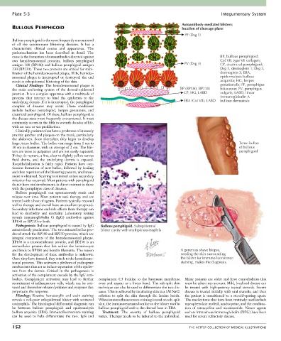
Plate 5-3                                                                                             Integumentary System

                                                                                          Autoantibody-mediated blisters:
       BULLOUS PEMPHIGOID                                                                 location of cleavage plane
                                                                                           PF (Dsg 1)
       Bullous pemphigoid is the most frequently encountered
       of  all  the  autoimmune  blistering  diseases.  It  has  a
       characteristic  clinical  course  and  appearance.  The
       pathomechanism  has  been  described  in  detail.  The
       cause is the formation of autoantibodies directed against                                            BP, bullous pemphigoid;
       two  hemidesmosomal  proteins,  bullous  pemphigoid                                                  Col VII, type VII collagen;
       antigen 180 (BP180) and bullous pemphigoid antigen                                  PV (Dsg 3)       CP, cicatricial pemphigoid;
       230 (BP230). These two proteins are critical for stabi-                                              Dsg 1, desmoglein 1; Dsg 3,
       lization of the hemidesmosomal plaque. If the hemides-                                               desmoglein 3; EBA,
       mosomal  plaque  is  interrupted  or  destroyed,  the  end                                           epidermolysis bullosa
       result is subepidermal blistering of the skin.                                                       acquisita; HG, herpes
         Clinical  Findings:  The  hemidesmosomal  plaque  is                                               gestationalis; PF, pemphigus
       the  main  anchoring  system  of  the  dermal-epidermal                           BP (BP180, BP230)   foliaceous; PV, pemphigus
       junction. It is a complex apparatus with a multitude of                             CP, HG, LABD     vulgaris; LABD, linear
       proteins  that  interact  to  bind  the  epidermis  to  the                                          immunoglobulin A
       underlying dermis. If it is interrupted, the pemphigoid                             EBA (Col VII), LABD  bullous dermatosis
       complex  of  diseases  may  occur.  These  conditions
       include  bullous  pemphigoid,  herpes  gestationis,  and
       cicatricial pemphigoid. Of these, bullous pemphigoid is
       the disease state most frequently encountered. It most
       commonly occurs in the fifth to seventh decades of life,
       with no race or sex predilection.
         Clinically, patients often have a prodrome of intensely
       pruritic patches and plaques on the trunk, particularly
       the  abdomen.  Soon  thereafter,  they  begin  to  develop
       large, tense bullae. The bullae can range from 1 cm to                                                        Tense bullae
       10 cm in diameter, with an average of 2 cm. The blis-                                                         of bullous
       ters are tense to palpation and are not easily ruptured.                                                      pemphigoid
       If they do rupture, a fine, clear to slightly yellow serous
       fluid  drains,  and  the  underlying  dermis  is  exposed.
       Reepithelialization  is  fairly  rapid.  Patients  have  con-
       tinuous formation of new bullae, followed by healing
       and then repetition of the blistering pattern, until treat-
       ment is obtained. Scarring is minimal unless secondary
       infection has occurred. Most patients with pemphigoid
       do not have oral involvement, in direct contrast to those
       with the pemphigus class of diseases.
         Bullous  pemphigoid  can  spontaneously  remit  and
       relapse over time. Most patients seek therapy and are
       treated with a host of agents. Patients typically respond
       well to therapy and overall have an excellent prognosis.
       Secondary infections and side effects from therapy can
       lead  to  morbidity  and  mortality.  Laboratory  testing
       reveals  immunoglobulin  G  (IgG)  antibodies  against
       BP180 or BP230 or both.
         Pathogenesis: Bullous pemphigoid is caused by IgG   Bullous pemphigoid. Subepidermal
       autoantibody production. The two autoantibodies pro-  blister cavity with multiple eosinophils
       duced attack the BP180 and BP230 proteins, which are
       integral  components  of  the  hemidesmosomal  plaque.
       BP180  is  a  transmembrane  protein,  and  BP230  is  an
       intracellular  protein  that  lies  within  the  keratinocyte
       and binds to BP180 and keratin filaments. The reason                              A generous shave biopsy,
       for  the  development  of  these  antibodies  is  unknown.                        sending the skin surrounding
       Once they have formed, they attach to the hemidesmo-                              the blister for immunofluorscence
       somal proteins. This activates a plethora of pathogenic                           staining, makes the diagnosis.
       mechanisms that act to induce separation of the epider-
       mis  from  the  dermis.  Critical  in  the  pathogenesis  is
       activation of the complement cascade by the IgG anti-
       bodies.  Complement  activation  may  lead  to  further   complement  C3  localize  to  the  basement  membrane   Many  patients  are  older  and  have  comorbidities  that
       recruitment of inflammatory cells, which can be acti-  zone  and  appear  as  a  linear  band.  The  salt-split  skin   must be taken into account. Mild, localized disease can
       vated and thereafter release cytokines and enzymes that   technique can also be used to differentiate the two dis-  be  treated  with  high-potency  topical  steroids.  Severe
       perpetuate the response.                  eases. This is achieved by incubating skin in a 1M NaCl   disease is treated initially with oral steroids, and then
         Histology:  Routine  hematoxylin  and  eosin  staining   solution  to  split  the  skin  through  the  lamina  lucida.   the  patient  is  transitioned  to  a  steroid-sparing  agent.
       reveals a cell-poor subepidermal blister with scattered   When immunofluorescence staining is used on salt-split   The medications that have been routinely used include
       eosinophils. The histological differential diagnosis can   skin, the immunoreactants localize to the blister roof in   mycophenolate mofetil, azathioprine, and the combina-
       be  between  bullous  pemphigoid  and  epidermolysis   bullous pemphigoid and to the dermal base in EBA.  tion  of  tetracycline  and  nicotinamide.  Newer  agents
       bullosa acquisita (EBA). Immunofluorescence staining   Treatment:  The  severity  of  bullous  pemphigoid   such as intravenous immunoglobulin (IVIG) have been
       can  be  used  to  help  differentiate  the  two.  IgG  and   varies. Therapy needs to be tailored to the individual.   used for severe refractory disease.

       152                                                                                   THE NETTER COLLECTION OF MEDICAL ILLUSTRATIONS