
Page 176 - The Netter Collection of Medical Illustrations - Integumentary System_ Volume 4 ( PDFDrive )
P. 176
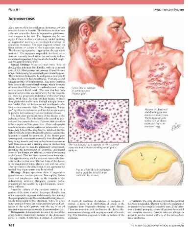
Plate 6-1                                                                                             Integumentary System


       ACTINOMYCOSIS


       Many species of the bacterial genus Actinomyces are able
       to cause disease in humans. The infection tends to run
       a chronic course that leads to suppurative granuloma-
       tous abscesses in the skin. The diagnosis may be sus-
       pected if there is clinical evidence of painful draining
       of  suppurative  material  and  histological  evidence  of
       granuloma formation. The exact diagnosis is based on
       tissue  culture  or  culture  of  the  suppurative  material.
       The disease is progressive if appropriate therapy is not
       instituted. The organisms responsible for these infec-
       tions are normally found within the oral cavity and are
       commensal organisms. They can also be found through-
       out the gastrointestinal tract.
         Clinical  Findings:  Males  are  much  more  likely  to
       develop this infection than females, with an estimated
       ratio of 3 : 1. Most patients are between 30 and 50 years
       of age. Predisposing factors include poor dental hygiene.
       The infection is believed to be endogenous in origin. It
       is a rare infection in the United States. There are several
       clinical pictures of actinomycosis. The most common
       form seen is the cervicofacial subtype, which accounts
       for more than 50% of cases. It is related to oral trauma,   Cervicofacial subtype
       such  as  recent  dental  work.  The  area  that  has  been   of actinomycosis
       traumatized provides a portal of entry for the bacteria,   (”lumpy jaw”)
       and there is a progressive induration of the underlying
       tissue.  With  time,  the  firm  swelling  begins  to  break
       through the skin and to drain through multiple cutane-
       ous fistulas. Pain can be intense and is relieved as the
       fistulas  spontaneously  drain.  The  designation  “lumpy
       jaw” signifies the induration and fistula formation seen                                        Abscess of chest wall
       in patients with actinomycosis cervicofacial disease.                                           and draining sinuses
         The next most prevalent form of the disease is the                                            due to actinomycosis.
       pulmonary form. This is believed to be caused by aspi-                                          The fungus spreads
       ration of the causative bacteria. Patients often complain                                       to the skin by direct
       of hemoptysis and low-grade fever. Chest radiographs                                            extension from the
       can show features similar to those of tuberculosis infec-                                       involved lung.
       tions. Any lobe of the lung may be involved, but the
       right lower lobe is most frequently affected because the
       infection  is  caused  by  aspiration.  If  the  disease  goes
       unrecognized, sinus tracts eventually form through the
       lung lining, muscle, and skin to the thoracic cutaneous
       wall. Skin abscess and a draining sinus in this location   The “ray fungus” as it appears in H&E-stained
       should  lead  one  to  look  for  pulmonary  involvement,   tissue section with surrounding neutrophilic
       including  the  development  of  empyema.  Abdominal   infiltrate
       forms of the disease are believed to occur after trauma
       to the bowel. This has been reported most frequently
       after appendectomy, and for unknown reasons the bac-
       teria localize to that area. The last form of the disease
       is the disseminated form, which is rare and can occur
       after any form of the disease that is not appropriately
       treated. Any organ system may be involved.      Pus in a Petri dish showing two
         Histology:  Biopsy  specimens  show  a  suppurative   sulfur granules (small lumps
       granulomatous  reaction  pattern.  Neutrophils,  histio-  indicated by arrows)
       cytes,  and  lymphocytes  make  up  the  majority  of  the
       inflammatory  infiltrate.  Basophilic  granules  (sulfur
       granules) are surrounded by a predominantly neutro-
       philic infiltrate.
         Anaerobic  culture  of  the  purulent  material  or  a
       portion of the tissue is critical for proper identification
       of the responsible organism and ultimately for choosing
       the appropriate therapy. Material should be sent anaero-
       bically immediately to the laboratory. Yellow to white   A.  meyeri,  A.  naeslundii,  A.  radingae,  A.  europaeus,  A.   Treatment: The drug of choice to treat this bacterial
       sulfur granules form as the culture material grows. Eval-  viscosus,  A.  neuii,  or  A.  odontolyticus.  A.  israelii  is  the   infection is penicillin. Therapy needs to be maintained
       uation  of  the  sulfur  granules  with  the  use  of  an  oil   organism  most  frequently  observed  to  cause  disease.   for months to be certain of complete cure. If the infec-
       immersion microscope shows the filamentous bacteria.  These are anaerobic, acid-fast bacteria that have a fila-  tion is treated promptly, almost all patients have a full
         Pathogenesis: Actinomycosis is caused by one of the   mentous morphology with varying amounts of branch-  and  complete  recovery.  Patients  who  are  allergic  to
       gram-positive  filamentous  bacteria  of  the  Actinomyces   ing. The definitive diagnosis is made by culture of the   penicillin can be treated with any of the tetracycline-
       genus: A. israelii, A. turicensis, A. lingnae, A. gravenitzii,   organism.          based antibiotics.

       162                                                                                   THE NETTER COLLECTION OF MEDICAL ILLUSTRATIONS