
Page 181 - The Netter Collection of Medical Illustrations - Integumentary System_ Volume 4 ( PDFDrive )
P. 181
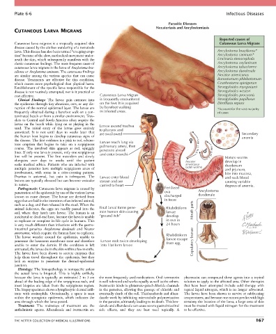
Plate 6-6                                                                                                Infectious Diseases

                                                                                     Parasitic Diseases
                                                                              Necatoriasis and Ancylostomiasis
        CUTANEOUS LARVA MIGRANS

                                                                                                              Reported causes of
        Cutaneous  larva  migrans  is  a  tropically  acquired  skin                                          Cutaneous Larva Migrans
        disease caused by the aimless wandering of a nematode
        larva. This disease has also been termed “creeping erup-                                             Ancylostoma braziliense*
        tion” because of the slow, methodical movement under-                                                Ancylostoma caninum*
        neath the skin, which subsequently manifests with the                                                Uncinaria stenocephala
        classic cutaneous findings. The most frequent cause of                                               Ancylostoma ceylanicum
        cutaneous larva migrans is the larva of Ancylostoma bra-                                             Ancylostoma tubaeforme
        ziliense or Ancylostoma caninum. The cutaneous findings                                              Ancylostoma duodenale
        are  similar  among  the  various  species  that  can  cause                                         Necator americanus
        disease.  Treatments  are  effective  for  this  condition,                                          Bunostomum phlebotomum
        which causes more psychological than physical harm.                                                  Gnathostoma spinigerum
        Establishment of the specific larva responsible for the                                              Strongyloides myopotami
        disease is not routinely attempted, nor is it practical or                                           Strongyloides westeri
        cost effective.                            Cutaneous Larva Migran                                    Strongyloides procyonis
          Clinical  Findings:  The  larvae  gain  entrance  into   is frequently encountered                 Strongyloides papillosus
        the epidermis through tiny abrasions, cuts, or any dis-  on the feet. It is acquired                 Dirofilaria repens
        ruption of the normal epidermal layer. The larvae are   by barefoot walking                          *Account for the vast majority
        frequently obtained during a barefoot walk on a con-  in infested areas.                             of cases
        taminated beach or from a similar environment. Trav-
        elers to Central and South America often acquire the
        larvae  on  the  beach  while  lying  on  or  playing  in  the   Larvae ascend trachea
        sand.  The  initial  entry  of  the  larvae  goes  entirely   to pharynx and
        unnoticed.  It  is  not  until  days  to  weeks  later  that     are swallowed                                   Secondary
        the human host begins to develop cutaneous signs of                                                              anemia
        the disease. The first evidence is a pink to red, edema-  Larvae reach lung via
        tous  eruption  that  begins  to  take  on  a  serpiginous   pulmonary artery, then
        course.  The  involved  skin  appears  as  red,  squiggly   penetrate alveoli              9 to 11 mm
        lines. If only one larva is present, only one serpiginous   and enter bronchi                    7 to 9 mm
        line  will  be  present.  The  line  meanders  and  slowly                                              Mature worms
        elongates  over  days  to  weeks  until  the  patient                                Necator            develop in
        seeks  medical  advice.  Patients  who  are  infected  with                          americanus         duodenum
        multiple  parasites  have  multiple  serpiginous  areas  of                          (adult worms)      and jejunum,
        involvement,  with  some  in  a  criss-crossing  pattern.                                               bite into mucosa,
        Pruritus  is  universal,  but  pain  is  infrequent.  The   Larvae enter blood                          and suck blood
        lesions are typically elevated but can become vesicular   stream and are                                causing variable
        in nature.                                carried to heart                                              degrees of anemia.
          Pathogenesis: Cutaneous larva migrans is caused by                       Fertilized
        penetration of the epidermis by one of the various larvae                  ova             Ancylostoma
                                                                                                   duodenale
        known  to  cause  disease.  The  larvae  are  derived  from                discharged
        eggs that are laid in the intestines of an infected animal,                in feces
        such as a dog, and then released in the stool. When the
        animal defecates, the eggs are readily passed into the   Final larval forms pene-  Rhabditiform  Mouth parts
        soil,  where  they  hatch  into  larvae.  The  human  is  an   trate human skin causing  larvae
        incidental or dead-end host, because the larva is unable   “ground itch”   develop
        to replicate or complete its life cycle in humans. This                    in ova in
        is very much different than infections with the gastro-                    24 hours
        intestinal  parasites  Ancylostoma  duodenale  and  Necator
        americanus, which require the human host to replicate.                     Rhabditiform
        The  larvae  wander  around  the  epidermis,  unable  to                   larvae escape
        penetrate the basement membrane zone and therefore   Larvae molt twice developing  from egg  Copulatory bursae
        unable  to  enter  the  dermis.  If  the  condition  is  left   into filariform larvae
        untreated, the larvae die in the skin within a few months.
        The  larvae  have  been  shown  to  secrete  enzymes  that
        help  them  travel  throughout  the  epidermis,  but  they
        lack  an  enzyme  to  penetrate  the  dermal-epidermal
        junction.
          Histology: The histopathology is nonspecific unless
        the  actual  larva  is  biopsied.  This  is  highly  unlikely,
        because  the  larva  is  typically  an  estimated  2  to  3 cm   the most frequently used medications. Oral ivermectin   pharmacist can compound these agents into a topical
        ahead of the leading edge of the serpiginous rash, and   is well tolerated and works equally as well as the others.   solution to apply to the affected area. Other therapies
        most  biopsies  are  taken  from  the  serpiginous  region.   Ivermectin binds to glutamate-gated chloride channels   that  have  been  attempted  include  cold  therapy  with
        The biopsy specimen shows a lymphocytic dermal infil-  in the parasites, allowing free passage of chloride and   topical liquid nitrogen, which is no longer advocated.
        trate  with  eosinophils.  Occasionally,  a  space  is  seen   eventually death of the cell. Thiabendazole and alben-  The larvae have been shown to survive at subfreezing
        within  the  spongiotic  epidermis,  which  indicates  the   dazole work by inhibiting microtubule polymerization   temperatures, and because one cannot predict with high
        area through which the larva passed.      in the parasite, ultimately leading to its death. Thiaben-  certainty the location of the larva, a large area of skin
          Treatment:  The  mainstays  of  treatment  are  the   dazole and albendazole can cause severe gastrointestinal   must be treated with liquid nitrogen for the treatment
        anthelmintic  agents.  Albendazole  and  ivermectin  are   side  effects,  and  they  are  best  used  topically.  A   to be effective.


        THE NETTER COLLECTION OF MEDICAL ILLUSTRATIONS                                                                          167