
Page 179 - The Netter Collection of Medical Illustrations - Integumentary System_ Volume 4 ( PDFDrive )
P. 179
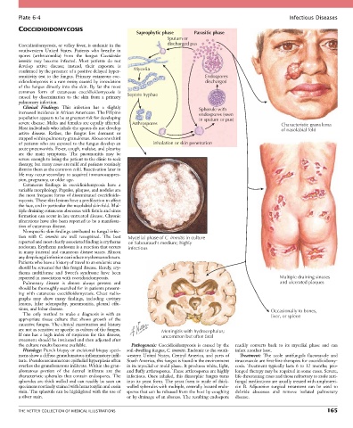
Plate 6-4                                                                                                Infectious Diseases
        COCCIDIOIDOMYCOSIS
                                                      Saprophytic phase     Parasitic phase
                                                                  Sputum or
        Coccidioidomycosis, or valley fever, is endemic in the    discharged pus
        southwestern  United  States.  Patients  who  breathe  in
        spores  (arthroconidia)  from  the  fungus  Coccidioides
        immitis  may  become  infected.  Most  patients  do  not
        develop  active  disease;  instead,  their  exposure  is
        confirmed by the presence of a positive delayed hyper-  Mycelia
        sensitivity test to the fungus. Primary cutaneous coc-                  Endospores
        cidioidomycosis is a rare entity caused by inoculation                  discharged
        of  the  fungus  directly  into  the  skin.  By  far  the  most
        common  form  of  cutaneous  coccidioidomycosis  is
        caused  by  dissemination  to  the  skin  from  a  primary   Septate hyphae
        pulmonary infection.
          Clinical  Findings:  This  infection  has  a  slightly              Spherule with
        increased incidence in African Americans. The Filipino                endospores (seen
        population appears to be at greatest risk for developing              in sputum or pus)
        severe disease. Males and females are equally affected.   Arthrospores                                Characteristic granuloma
        Most individuals who inhale the spores do not develop                                                 of nasolabial fold
        active  disease.  Rather,  the  fungus  lies  dormant  or
        trapped within pulmonary granulomas. About one third
        of patients who are exposed to the fungus develop an   Inhalation or skin penetration
        acute pneumonitis. Fever, cough, malaise, and pleurisy
        are  the  main  symptoms.  The  pneumonitis  may  be
        severe enough to bring the patient to the clinic to seek
        therapy, but many cases are mild and patients routinely
        dismiss them as the common cold. Reactivation later in
        life may occur secondary to acquired immunosuppres-
        sion, pregnancy, or older age.
          Cutaneous  findings  in  coccidioidomycosis  have  a
        variable morphology. Papules, plaques, and nodules are
        the most frequent forms of disseminated coccidioido-
        mycosis. These skin lesions have a predilection to affect
        the face, and in particular the nasolabial skin fold. Mul-
        tiple draining cutaneous abscesses with fistula and sinus
        formation can occur in late untreated disease. Chronic
        ulcerations have also been reported to be a manifesta-
        tion of cutaneous disease.
          Nonspecific skin findings attributed to fungal infec-
        tion  with  C.  immitis  are  well  recognized.  The  best   Mycelial phase of C. immitis in culture
        reported and most clearly associated finding is erythema   on Sabouraud’s medium; highly
        nodosum. Erythema nodosum is a reaction that occurs   infectious
        in many internal and cutaneous disease states. Almost
        any deep fungal infection can induce erythema nodosum.
        Patients who have a history of travel to an endemic area
        should be screened for this fungal disease. Rarely, ery-
        thema  multiforme  and  Sweet’s  syndrome  have  been
        reported in association with coccidioidomycosis.                                                     Multiple draining sinuses
          Pulmonary  disease  is  almost  always  present  and                                               and ulcerated plaques
        should be thoroughly searched for in patients present-
        ing  with  cutaneous  coccidioidomycosis.  Chest  radio-
        graphs  may  show  many  findings,  including  cavitary
        lesions,  hilar  adenopathy,  pneumonitis,  pleural  effu-
        sions, and lobar disease.                                                                         Occasionally to bones,
          The  only  method  to  make  a  diagnosis  is  with  an                                         liver, or spleen
        appropriate  tissue  culture  that  shows  growth  of  the
        causative fungus. The clinical examination and history
        are not as sensitive or specific as culture of the fungus.   Meningitis with hydrocephalus;
        If  one  has  a  high  index  of  suspicion  for  this  disease,   uncommon but often fatal
        treatment should be instituted and then adjusted after
        the culture results become available.       Pathogenesis:  Coccidioidomycosis  is  caused  by  the   readily  converts  back  to  its  mycelial  phase  and  can
          Histology: Punch biopsy or excisional biopsy speci-  soil-dwelling fungus, C. immitis. Endemic to the south-  infect another host.
        mens show a diffuse granulomatous inflammatory infil-  western United States, Central America, and parts of   Treatment:  The  azole  antifungals  fluconazole  and
        trate. Pseudocarcinomatous epithelial hyperplasia often   South America, this fungus is found in the environment   itraconazole are first-line therapies for coccidioidomy-
        overlies the granulomatous infiltrate. Within the gran-  in its mycelial or mold phase. It produces white, light,   cosis.  Treatment  typically  lasts  6  to  12  months;  pro-
        ulomatous  portion  of  the  dermal  infiltrate  are  the     and fluffy arthrospores. These arthrospores are highly   longed therapy may be required in some cases. Severe,
        characteristic  spherules  that  contain  endospores.  The   infectious. Once inhaled, this dimorphic fungus turns   life-threatening cases and those refractory to azole anti-
        spherules are thick walled and can readily be seen on   into its yeast form. The yeast form is made of thick-  fungal medications are usually treated with amphoteri-
        specimens routinely stained with hematoxylin and eosin   walled spherules with multiple, centrally located endo-  cin  B.  Adjunctive  surgical  treatment  can  be  used  to
        stain. The spherule can be highlighted with the use of   spores that can be released from the host by coughing   debride  abscesses  and  remove  isolated  pulmonary
        a silver stain.                           or by drainage of an abscess. The resulting endospore   disease.


        THE NETTER COLLECTION OF MEDICAL ILLUSTRATIONS                                                                          165