
Page 183 - The Netter Collection of Medical Illustrations - Integumentary System_ Volume 4 ( PDFDrive )
P. 183
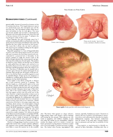
Plate 6-8                                                                                                Infectious Diseases

                                                                               TINEA CRURIS AND TINEA CAPITIS


        DERMATOPHYTOSES (Continued)


        general public, because of personal involvement or that
        of someone they know. This fungal infection is seen in
        two  predominant  types,  the  interdigital  type  and  the
        moccasin type. The interdigital subtype forms macer-
        ated,  red  patches  in  the  toe  web  spaces.  The  areas
        can  become  pruritic  and  can  lead  to  onychomycosis.
        Moccasin-type tinea pedis involves the entire foot and
        is the less common of the two types. T. rubrum is the
        most frequent isolate in these cases.
          Tinea  manuum,  also  most  frequently  caused  by  T.
        rubrum, predominantly affects one hand only. It is com-                                       Tinea cruris (male), “jock itch,”
        monly seen in association with bilateral tinea pedis and   Tinea cruris (female)              a very common infecton in males
        therefore has been called “one hand two feet disease.”
        The reason that it affects only one hand is unknown.
        The most frequent complaint is itching and the appear-
        ance of the red annular patches.
          Majocchi’s granuloma is a form of fungal folliculitis
        caused by one of the dermatophyte species. It is univer-
        sally seen in patients who have been treated with corti-
        costeroids  for  a  presumed  form  of  dermatitis.  As  the
        patient  continues  to  apply  the  steroid  cream  to  the
        patch of fungal infection, the redness spreads, and pus-
        tules may form within the affected region. The pustules
        are based on a hair follicle, and the hair may be absent
        or  easily  pulled  from  the  region  with  minimal  or  no
        discomfort. Removal of the hair and use of a potassium
        hydroxide (KOH) preparation allows the fungus to be
        seen.  This  form  of  folliculitis  must  be  treated  with  a
        systemic agent, because the topical antifungals do not
        penetrate deep enough into the depths of the hair fol-
        licle or into the hair shaft, as would be required to treat
        an endothrix fungus. Fungal species are designated as
        endothrix or ectothrix species based on their ability to
        penetrate the hair shaft epithelium.
          Tinea  capitis  is  seen  almost  exclusively  in  children
        and  is  most  commonly  caused  by  T.  tonsurans.  This
        infection begins as a small, pruritic patch in the scalp
        that  slowly  expands  outward.  Hair  loss  is  prominent
        because the fungus invades the hair shaft and can cause
        the hair to break. A frequent clinical sign is “black dot”
        tinea.  This  is  the  clinical  finding  of  tiny,  broken-off
        hairs that appear as black dots just at the level of the
        scalp. Posterior occipital adenopathy is always seen in
        cases of tinea capitis, and its absence should make one
        reconsider the diagnosis. If a child presents with a scaly
        patch in the scalp and associated hair loss, it should be
        treated as tinea capitis until proven otherwise. A KOH
        examination of the hair or of a scalp scraping often, but
        not always, shows evidence of a dermatophyte. A fungal
        culture can be used in these cases to confirm the diag-
        nosis if the KOH examination is negative. The culture        Tinea capitis. Scaly patches with associated alopecia
        sample  is  easily  obtained  by  rubbing  the  scaly  patch
        with  a  toothbrush  and  collecting  the  scale  that  is
        removed in a sterile container. The cultures are grown
        in the laboratory on dermatophyte test medium (DTM),   class.  The  kerion  often  appears  as  a  large,  inflamed,   bacteria, especially Staphylococcus species. Treatment is
        and growth is often seen in 2 to 4 weeks. Tinea capitis   boggy-feeling  plaque  with  alopecia.  Serous  drainage   based on the use of systemic oral antifungals in associa-
        requires  at  least  6  weeks  of  systemic  oral  therapy  to   and crusting are also present. These plaques are very   tion with an oral corticosteroid to decrease the massive
        clear, and all the patients’ pets, especially cats, should   tender  to  palpation,  and  children  complain  of  pain     inflammatory  response.  Any  bacterial  coinfections
        be evaluated by a veterinarian for evidence of disease.  even  when  the  lesions  are  not  manipulated.  Alopecia   must  be  treated  at  the  same  time.  Scarring  alopecia
          A kerion is a boggy plaque found on occasion in tinea   overlies  the  plaque,  and  if  it  is  severe,  a  kerion  can     may be permanent and may lead to morbidity for the
        capitis that results from a massive immune inflamma-  lead to permanent scarring alopecia. Posterior occipital   child.
        tory response to the causative fungal agent. The fungi   and cervical adenopathy is present and tender to palpa-  Tinea unguium, or onychomycosis, is clinically rec-
        most likely to cause this reaction are in the zoophilic   tion.  The  kerion  often  become  impetiginized  with   ognized by thick, dystrophic, crumbling nails. One or


        THE NETTER COLLECTION OF MEDICAL ILLUSTRATIONS                                                                          169