
Page 184 - The Netter Collection of Medical Illustrations - Integumentary System_ Volume 4 ( PDFDrive )
P. 184
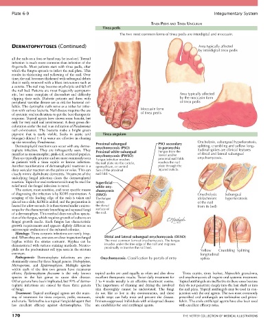
Plate 6-9                                                                                             Integumentary System

                                                                             TINEA PEDIS AND TINEA UNGUIUM
                                                     Tinea pedis
                                                             The two most common forms of tinea pedis are interdigital and moccasin.


       DERMATOPHYTOSES (Continued)                                                                   Area typically affected
                                                                                                     by interdigital tinea pedis

       all the nails on a foot or hand may be involved. Toenail
       infection is much more common than infection of the
       fingernails.  Most  patients  start  with  tinea  pedis,  after
       which the fungus spreads to infect the nail plate. This
       results  in  thickening  and  yellowing  of  the  nail.  Over
       time, the nail becomes thickened with subungual debris
       that is easily removed with a blunt instrument such as
       a curette. The nail may become onycholytic and fall off
       the nail bed. Patients are most frequently asymptom-
       atic,  but  some  complain  of  discomfort  and  difficulty                         Area typically affected
       clipping  their  nails.  Diabetic  patients  and  those  with                       by the moccasin form
       peripheral vascular disease are at risk for bacterial cel-                          of tinea pedis
       lulitis. The dystrophic nails serve as a nidus for infec-
       tion with various bacteria. Nail disease requires the use        Moccasin form
       of systemic oral medications to get the best therapeutic         of tinea pedis
       response. Topical agents have shown some benefit, but
       only for very mild nail involvement. A deep green dis-
       coloration under the nail is an indication of Pseudomonas
       nail  colonization.  The  bacteria  make  a  bright  green
       pigment  that  is  easily  visible.  Soaks  in  acetic  acid   Tinea unguium
       (vinegar) diluted 1 : 4 in water are effective in clearing
       up the secondary Pseudomonas.             Proximal subungual              PSO secondary      Oncholysis, subungual hyperkeratosis,
         Dermatophytid reactions can occur with any derma-                                          splitting, crumbling and yellow long-
       tophyte  infection.  They  are  infrequently  seen.  They   onychomycosis (PSO)  to paronychia  itudinal spikes are clinical features
                                                                                 Fungus from the
                                                 Proximal white subungual
       manifest as monomorphic, pink-red, scattered papules.   onychomycosis (PWSO)  lateral and/or  of distal and lateral subungual
       They are typically pruritic and are most commonly seen   Fungus infection reaches  proximal nail folds  onychomycosis.
       in  patients  with  a  tinea  capitis  or  kerion  infection.   the nail plate via the cuticle,  reaches the nail
       Another manifestation of dermatophytid reactions is a   eponychium, or ventral  plate through the
       deep vesicular reaction on the palms or soles. This can   face of the proximal  injured cuticle.
       closely mimic dyshidrotic dermatitis. Treatment of the   nail fold.
       underlying  fungal  infection  clears  the  dermatophytid
       reaction. Topical or oral corticosteroids may be used for   Superficial
       relief until the fungal infection is cured.  white ony-
         The easiest, most sensitive, and most specific means   chomycosis
       of diagnosing the infection is by KOH examination. A   (SWO)                                 Onycholysis   Subungual
       scraping of the leading edge of the rash is taken and   The fungus                           (detachment   hyperkeratosis
       placed on a slide; KOH is added, and the preparation is   infects                            of the nail
       heated for a few seconds. It is then viewed under a micro-  the dorsal                       from its bed)
       scope for the characteristic branching and septated fungi   surface of
       of a dermatophyte. This method does not allow specia-  the nail.
       tion of the fungus, which requires growth of cultures on
       fungal  growth  media.  Each  fungus  has  characteristic
       growth requirements and appears slightly different on
       microscopic evaluation of the cultured colonies.
         Histology: Tinea corporis infections are rarely biop-
       sied. When they are, one sees on close inspection fungal   Distal and lateral subungual onychomycosis (DLSO)
       hyphae  within  the  stratus  corneum.  Hyphae  can  be   The most common form of onychomycosis. The fungus
       demonstrated with various staining methods. Neutro-  invades under the free edge of the nail and migrates
       phils are the predominant cell type seen in the stratum   proximally to involve the nail bed.  Yellow   Crumbling Splitting
       corneum.                                                                                       longitudinal
         Pathogenesis:  Dermatophyte  infections  are  pre-  Onychomycosis. Classification by portals of entry  spikes
       dominantly caused by three fungal genera: Trichophyton,
       Microsporum,  and  Epidermophyton.  Multiple  species
       within  each  of  the  first  two  genera  have  cutaneous
       effects;  Epidermophyton  floccosum  is  the  only  known   topical azoles are used equally as often and also show   Tinea  capitis,  tinea  barbae,  Majocchi’s  granuloma,
       species  in  the  last  genus  to  cause  skin  disease.     excellent therapeutic results. Twice-daily treatment for   and onychomycosis all require oral systemic treatment.
       Other genera have been implicated, but 99% of derma-  2 to 4 weeks usually is an effective treatment course.   Topical antifungals are ineffective in these cases because
       tophyte  infections  are  caused  by  these  three  genera     The  importance  of  cleaning  and  drying  the  involved   they do not penetrate deeply into the hair shaft or into
       of fungi.                                 skin  thoroughly  cannot  be  understated.  The  fungi     the nail plate. Topical antifungals may be used in con-
         Treatment: Topical antifungal agents are the main-  do  not  like  to  live  in  dry  environments,  and  these     junction with the oral agents. The two most commonly
       stay  of  treatment  for  tinea  corporis,  pedis,  manuum,   simple  steps  can  help  treat  and  prevent  the  disease.   prescribed oral antifungals are terbinafine and griseo-
       and cruris. Terbinafine is a topical fungicidal agent that   Immunosuppressed individuals with widespread disease   fulvin. The azole antifungal agents have also been used
       has  excellent  efficacy  against  dermatophytes.  The   are candidates for oral antifungal agents.  with excellent efficacy rates.

       170                                                                                   THE NETTER COLLECTION OF MEDICAL ILLUSTRATIONS