
Page 208 - The Netter Collection of Medical Illustrations - Integumentary System_ Volume 4 ( PDFDrive )
P. 208
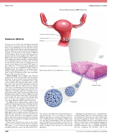
Plate 6-33                                                                                            Integumentary System

                                                                         HUMAN PAPILLOMAVIRUS (HPV) INFECTION


                                                 Uterine cervical canal

       VERRUCAE (WARTS)                          Uterine cervix
                                                 Vagina

       Verrucae are one of the most frequently encountered
       viral infections in humans. They are capable of causing
       disease in any individual, but severe infections seem to
       be more likely in those who are immunocompromised.
       Warts can affect any cutaneous surface, and unique wart
       subtypes are more prone to cause disease in different
       clinical locations. By far the most important aspect of
       infection with the human papillomavirus (HPV) is the                                                              Vaginal
       ability of the virus to cause malignant transformation.                                                           lumen
       This malignant potential is specific to certain subtypes
       and is especially a concern in women, who are at risk
       for cervical cancer. Most cases of cervical cancer can be
       traced to prior infection with certain HPV strains. In
       June  2006,  the  U.S.  Food  and  Drug  Administration   Stratified cervical epithelium
       approved  the  use  of  a  prophylactic  HPV  vaccine  in
       prepubertal girls. The vaccine is a recombinant quad-
       rivalent vaccine against HPV types 6, 11, 16, and 18.   Basal layer of the cervical epithelium
       Types 16 and 18 are believed to have been responsible
       for up to 70% of cervical cancers.
         Clinical  Findings:  Verruca  vulgaris,  also  called  the
       common wart, is the most prevalent wart that infects
       the human. It can be located on any cutaneous surface.
       These warts often appear as small papules with a rough
       surface  studded  with  pinpoint,  dark  purple  to  black
       dots. These dots represent the thromboses of the tiny
       capillaries  within  the  wart.  Most  warts  are  between                                               Lamina propria
       5 mm and 1 cm in diameter, but some can become quite   Virus
       large and encompass much larger areas of the skin. The
       coalescence  of  multiple  warts  into  one  larger  wart  is
       called mosaic warts. These are most commonly seen on
       the plantar aspect of the foot. Verruca vulgaris can come
       in many sizes and shapes. Most lesions spontaneously
       resolve within a few years. A good rule of thumb is that
       50%  of  verrucae  will  disappear  spontaneously  in  2
       years. Many distinctive clinical forms of warts exist.
         The filiform wart is represented by a small verrucal
       papule with finger-like projections extruding from the
       base of the papule. The projections are typically 1 to
       2 mm thick and 4 to 7 mm long. They are commonly
       found on the face. The flat wart is frequently encoun-
       tered and manifests as a 3- to 5-mm, flat papule with a
       slight pink to red to purple coloration. Flat warts are
       frequently seen on the legs of women and in the beard   The warts are well defined and characteristically inter-  Subungual and periungual warts, a subclassification
       region  of  men,  and  they  can  be  arranged  in  a  linear   rupt  the  skin  lines.  This  is  in  contrast  to  a  callus,  in   of  palmar/plantar  warts,  are  found  around  and  under
       pattern if the warts are spread during the act of shaving.   which the skin lines are retained, and this sign can be   the nail apparatus. These warts can cause nail dystrophy
       Flat warts have been found to be highly associated with   used to differentiate the two conditions. Plantar warts   and  pain  on  grasping  of  objects.  They  tend  to  affect
       HPV types 3 and 10.                       can  cause  pain  and  discomfort  if  they  are  located  in   more than one finger and can be more difficult to treat
         Plantar  warts  (myrmecia)  are  seen  on  the  plantar   areas of pressure such as the heels or across the skin   than the common wart. Long-standing periungual or
       aspect of the foot and are caused for the most part by   underlying the metatarsal heads. Palmar warts are very   subungual  warts  that  have  a  changing  morphology
       HPV types 1, 2, and 4. They are deep-seated papules   similar  to  plantar  warts  and  have  the  same  clinical   should be biopsied to rule out malignant transforma-
       and plaques that may coalesce into large mosaic warts.   appearance.                tion  into  a  squamous  cell  carcinoma.  This  is  not

       194                                                                                   THE NETTER COLLECTION OF MEDICAL ILLUSTRATIONS