
Page 204 - The Netter Collection of Medical Illustrations - Integumentary System_ Volume 4 ( PDFDrive )
P. 204
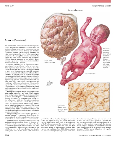
Plate 6-29                                                                                            Integumentary System

                                                                                SYPHILIS IN PREGNANCY


       SYPHILIS (Continued)


       and tabes dorsalis. Tabes dorsalis results from degenera-
       tion of the posterior columns of the spinal cord. The
       posterior columns are critical for proper sensation, and                                                           Macerated
       patients  with  tabes  dorsalis  develop  gait  disorders,                                                         erosions,
       diminished  reflexes,  proprioception  abnormalities,                                                              sloughed
       pain,  paresthesias,  and  a  host  of  other  neurological                                                        skin, failure
       symptoms.  If  neurosyphilis  remains  untreated,  the                                                             to thrive.
       patient dies of the disease. Therefore, any patient who                                                            Approx-
       exhibits  signs  or  symptoms  of  neurosyphilis  should                                                           imately
       undergo a spinal tap to evaluate the cerebrospinal fluid   Large, pale,                                            1/3 of neo-
       for involvement with T. pallidum.             boggy placenta                                                       nates will
         Congenital  syphilis  occurs  as  the  result  of  vertical                                                      die from
       transmission  from  an  infected  mother  to  her  unborn                                                          congenital
       fetus. Up to one third of infected neonates die of the                                                             syphilis.
       disease. In neonates who survive, the disease manifests
       in many ways. Neonates may present with macerated
       erosions associated with cachexia and failure to thrive.                                     Macerated fetus
       “Snuffles”  is  the  term  used  to  describe  the  chronic
       runny nose with a bloody purulent discharge. Rhagades
       are one of the most common signs seen in congenital
       syphilis; they appear as scarring around the mouth and
       eyes.  Many  bony  abnormalities  have  been  reported,
       including a saddle-nose deformity, the Higoumenakis
       sign  (medial  clavicular  thickening),  saber  shins,  and
       Clutton’s joints. Teeth abnormalities include Hutchin-
       son’s teeth (notched incisors) and, less frequently, mul-
       berry molars.
         Histology: Skin biopsies of syphilis that are evaluated
       with  routine  hematoxylin  and  eosin  (H&E)  staining
       show varying features depending on the stage and form
       of  disease  being  biopsied.  A  universal  finding  in  all
       forms is the presence of numerous plasma cells within
       the  inflammatory  infiltrate.  Ulceration,  granulomas,
       and vasculitis are often encountered. The spirochetes
       cannot  be  appreciated  with  routine  H&E  staining;
       special  staining  techniques  are  required.  The  Steiner   Spirochetes
       stain  and  the  Warthin-Starry  stain  are  the  two  most   in fetal tissue
       commonly  used  stains.  Immunohistochemical  stains         (Levaditi stain).
       can also be used, and they have been shown to be highly      T. pallidum
       sensitive and specific.
         Pathogenesis: Syphilis is caused by the spirochete, T.
       pallidum pallidum. This bacteria is highly infective and
       is predominantly spread by sexual contact and by trans-
       mission from an infected mother to her unborn child.  penicillin for at least 2 weeks. Most patients who are   the rash of secondary syphilis appear worse for a period
         Treatment: The T. pallidum organism has very little   treated  for  syphilis  develop  the  Jarisch-Herxheimer   of time. This reaction is not specific to T. pallidum and
       antibiotic resistance, and the therapy of choice is still   reaction. This reaction is the result of the decimation   has  been  reported  with  other  infectious  agents.  It  is
       penicillin. A single intramuscular dose of 2.4 million IU   of the T. pallidum organisms due to therapy with peni-  critical  to  follow  patients  long  enough  after  therapy
       of benzathine penicillin G is recommended, and some   cillin.  As  the  scores  of  bacteria  are  killed,  the  dead   ensure  adequate  treatment  as  measured  by  titers  on
       now  recommend  a  follow-up  dose—the  same  as  the   spirochetes  induce  an  inflammatory  reaction.  This   rapid plasma reagin (RPR) or venereal disease research
       initial  dose—at  1  or  2  weeks.  Patients  who  develop   reaction may manifest as fever, chills, fatigue, malaise,   laboratory (VDRL) testing. All patients with syphillis
       neurosyphilis  need  to  be  treated  with  intravenous   and rashes of varying morphology. It can often make   should be tested for HIV.

       190                                                                                   THE NETTER COLLECTION OF MEDICAL ILLUSTRATIONS