
Page 214 - The Netter Collection of Medical Illustrations - Integumentary System_ Volume 4 ( PDFDrive )
P. 214
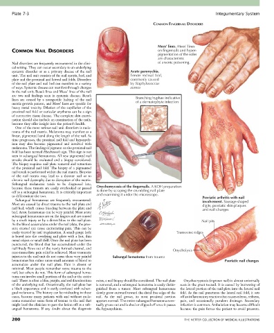
Plate 7-3                                                                                             Integumentary System

                                                                              COMMON FINGERNAIL DISORDERS


                                                                                Mees’ lines. Mees’ lines
       COMMON NAIL DISORDERS                                                    on fingernails and hyper-
                                                                                pigmentation of the soles
                                                                                are characteristic
       Nail disorders are frequently encountered in the clini-                  of arsenic poisoning.
       cal setting. They can occur secondary to an underlying
       systemic  disorder  or  as  a  primary  disease  of  the  nail   Acute paronychia.
       unit. The nail unit consists of the nail matrix, bed, and   Tender red nail fold,
       plate and the proximal and lateral nail folds. Disorders   commonly caused
       of the nail plate and nail bed can manifest in a variety   by Staphylococcus
       of ways. Systemic disease can manifest through changes     aureus
       in the nail unit. Beau’s lines and Mees’ lines of the nail
       are  two  nail  findings  seen  in  systemic  disease.  Beau’s
       lines  are  caused  by  a  nonspecific  halting  of  the  nail   Branching hyphae indicative
       matrix growth pattern, and Mees’ lines are specific for       of a dermatophyte infection
       heavy metal toxicity. Dilation of the capillaries of the
       proximal nail fold or cuticular erythema can be a sign
       of connective tissue disease. The complete skin exami-
       nation should also include an examination of the nails,
       because they offer insight into the patient’s health.
         One of the most serious nail unit disorders is mela-
       noma of the nail matrix. Melanoma may manifest as a
       linear, pigmented band along the length of the nail. As
       time progresses, the proximal nail fold and hyponych-
       ium  may  also  become  pigmented  and  involved  with
       melanoma. The finding of pigment on the proximal nail
       fold has been termed Hutchinson’s sign. This sign is not
       seen in subungual hematomas. All new pigmented nail
       streaks  should  be  evaluated  and  a  biopsy  considered.
       The biopsy requires nail plate removal and retraction
       of the proximal nail fold. The biopsy of a pigmented
       nail streak is performed within the nail matrix. Biopsies
       of  the  nail  matrix  may  lead  to  a  thinner  nail  or  to
       chronic nail dystrophy due to disruption of the matrix.
       Subungual  melanoma  tends  to  be  diagnosed  late,
       because  these  tumors  are  easily  overlooked  or  passed   Onychomycosis of the fingernails. A KOH preparation
       off as a subungual hematoma. It is critically important   is done by scraping the crumbling nail plate
       to differentiate the two.                 and examining it under the microscope.               Psoriatic arthritis with nail
         Subungual  hematomas  are  frequently  encountered.                                          involvement. Sausage-shaped
       Most are caused by direct trauma to the nail plate and                                         digits, psoriatic skin plaques
       nail bed, which causes bleeding between the plate and                                          and nail changes
       bed. Acute hematomas can be very painful. Most acute
       subungual hematomas are on the fingers and are caused
       by a crush injury or by a direct blow to the nail plate.                                       Nail pits
       As the blood accumulates under the nail plate, the pres-
       sure created can cause excruciating pain. This can be
       easily treated by nail trephination. A small-gauge hole                           Transverse ridges
       is bored into the overlying nail plate with a hot, thin
       metal object or small drill. Once the nail plate has been
       punctured, the blood that has accumulated under the
       nail freely flows out of the newly formed channel, and                          Onycholysis
       near-immediate pain relief is achieved. Most traumatic
       injuries to the nail unit do not cause these very painful   Subungal hematoma from trauma
       hematomas but rather cause small amounts of blood to                                                      Psoriatic nail changes
       accumulate  under  the  nail  plate.  Pain  is  absent  or
       minimal. Most people remember some trauma to the
       nail, but others do not. This form of subungual hema-
       toma can involve small portions of the nail or the entire
       nail. There is often a blue, purple, and red discoloration   exists, a nail biopsy should be considered. The nail plate   Onychocryptosis (ingrown nail) is almost universally
       of the underlying nail. Occasionally, the nail plate has   is removed, and a subungual hematoma is easily distin-  seen in the great toenail. It is caused by burrowing of
       a black appearance and is easily confused with subun-  guished  from  a  tumor.  Most  subungual  hematomas   the lateral portion of the nail plate into the lateral nail
       gual melanoma. The history can be misleading in these   slowly grow outward toward the distal free edge of the   fold. As the nail punctures the lateral nail fold, it sets
       cases,  because  many  patients  with  and  without  mela-  nail.  As  the  nail  grows,  its  most  proximal  portion   off an inflammatory reaction that causes edema, redness,
       noma remember some form of trauma to the nail that   appears normal. The entire subungual hematoma even-  pain,  and  occasionally  purulent  drainage.  Secondary
       might lead the clinician to pass the lesion off as a sub-  tually grows out and is shed or clipped off once it passes   infection is common. Ambulation may become difficult
       ungual  hematoma.  If  any  doubt  about  the  diagnosis   the hyponychium.         because the pain forces the patient to avoid pressure.

       200                                                                                   THE NETTER COLLECTION OF MEDICAL ILLUSTRATIONS