
Page 219 - The Netter Collection of Medical Illustrations - Integumentary System_ Volume 4 ( PDFDrive )
P. 219
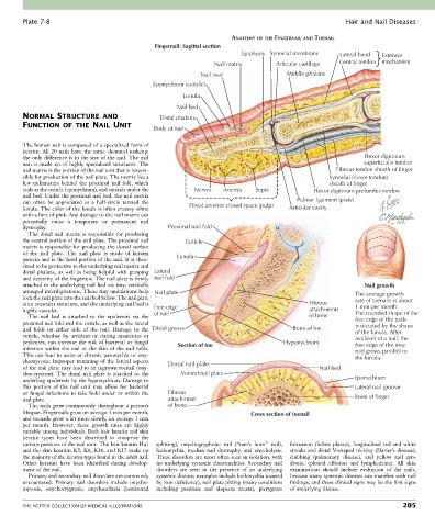
Plate 7-8                                                                                             Hair and Nail Diseases

                                                                          ANATOMY OF THE FINGERNAIL AND TOENAIL
                                                  Fingernail: Sagittal section
                                                                             Epiphysis Synovial membrane    Lateral band  Extensor
                                                                    Nail matrix         Articular cartilage  Central tendon  mechanism
                                                                Nail root                  Middle phalanx
                                                 Eponychium (cuticle)
                                                           Lunula
                                                         Nail bed
        NORMAL STRUCTURE AND                       Distal phalanx
        FUNCTION OF THE NAIL UNIT
                                                 Body of nail

        The human nail is composed of a specialized form of
        keratin. All 20 nails have the same chemical makeup;
        the only difference is in the size of the nail. The nail                                                   Flexor digitorum
        unit  is  made  up  of  highly  specialized  structures.  The                                              superficialis tendon
        nail matrix is the portion of the nail unit that is respon-                                        Fibrous tendon sheath of finger
        sible for production of the nail plate. The matrix lies a                                        Synovial (flexor tendon)
        few  millimeters  behind  the  proximal  nail  fold,  which                                      sheath of finger
        ends as the cuticle (eponychium), and extends under the   Nerves  Arteries  Septa           Flexor digitorum profundus tendon
        nail bed. Under the proximal nail bed, the nail matrix
        can  often  be  appreciated  as  a  half-circle  termed  the   Distal anterior closed space (pulp)  Palmar ligament (plate)
        lunula. The color of the lunula is often creamy white                               Articular cavity
        with a hint of pink. Any damage to the nail matrix can
        potentially  cause  a  temporary  or  permanent  nail
        dystrophy.                                    Proximal nail fold
          The  distal  nail  matrix  is  responsible  for  producing
        the ventral portion of the nail plate. The proximal nail   Cuticle
        matrix is responsible for producing the dorsal surface
        of  the  nail  plate.  The  nail  plate  is  made  of  keratin   Lunula
        protein and is the hard portion of the nail. It is theo-
        rized to be protective to the underlying nail matrix and
        distal  phalanx,  as  well  as  being  helpful  with  grasping  Lateral
        and dexterity of the fingertips. The nail plate is firmly  nail fold
        attached to the underlying nail bed via tiny, vertically                                                    Nail growth
        arranged interdigitations. These tiny undulations help  Nail plate                                       The average growth
        lock the nail plate into the nail bed below. The nail plate                                              rate of toenails is about
        is an avascular structure, and the underlying nail bed is   Free edge                     Fibrous        1 mm per month.
        highly vascular.                         of nail                                          attachments    The rounded shape of the
          The  nail  bed  is  attached  to  the  epidermis  via  the                              of bone        free edge of the nails
        proximal nail fold and the cuticle, as well as the lateral                                               is dictated by the shape
        nail  folds  on  either  side  of  the  nail.  Damage  to  the  Distal groove        Bone of toe         of the lunula. After
        cuticle,  whether  by  accident  or  during  manicures  or                                               avulsion of a nail, the
        pedicures, can increase the risk of bacterial or fungal   Section of toe          Hyponychium            free edge of the new
        infection within the nail or the skin of the nail folds.                                                 nail grows parallel to
        This can lead to acute or chronic paronychia or ony-                                                     the lunula.
        chomycosis. Improper trimming of the lateral aspects   Dorsal nail plate
        of the nail plate may lead to an ingrown toenail (ony-                                       Nail bed
        chocryptosis).  The  distal  nail  plate  is  attached  to  the   Ventral nail plate
        underling epidermis by the hyponychium. Damage to                                                       Eponychium
        this  portion  of  the  nail  unit  may  allow  for  bacterial                                          Lateral nail groove
        or fungal infections to take hold under or within the   Fibrous
        nail plate.                                   attachment                                                Bone of finger
          The nails grow continuously throughout a person’s   of bone
        lifespan. Fingernails grow on average 3 mm per month,                   Cross section of toenail
        and toenails grow a bit more slowly, on average 1 mm
        per  month.  However,  these  growth  rates  are  highly
        variable among individuals. Both hair keratin and skin
        keratin  types  have  been  described  to  comprise  the
        various portions of the nail unit. The hair keratin Ha1   splitting),  onychogryphotic  nail  (“ram’s  horn”  nail),   formation  (lichen  planus),  longitudinal  red  and  white
        and the skin keratins K5, K6, K16, and K17 make up   leukonychia,  median  nail  dystrophy,  and  onycholysis.   streaks and distal V-shaped nicking (Darier’s disease),
        the majority of the keratin types found in the adult nail.   These disorders are most often seen in isolation, with   clubbing  (pulmonary  disease),  and  yellow  nail  syn-
        Other  keratins  have  been  identified  during  develop-  no  underlying  systemic  abnormalities.  Secondary  nail   drome  (pleural  effusion  and  lymphedema).  All  skin
        ment of the nail.                         disorders  are  seen  in  the  presence  of  an  underlying   examinations  should  include  evaluation  of  the  nails,
          Primary and secondary nail disorders are commonly   systemic disease; examples include koilonychia (caused   because many systemic diseases can manifest with nail
        encountered.  Primary  nail  disorders  include  onycho-  by iron deficiency), nail plate pitting (many conditions   findings, and these clinical signs may be the first signs
        mycosis,  onychocryptosis,  onychoschizia  (horizontal   including  psoriasis  and  alopecia  areata),  pterygium   of underlying disease.


        THE NETTER COLLECTION OF MEDICAL ILLUSTRATIONS                                                                          205