
Page 215 - The Netter Collection of Medical Illustrations - Integumentary System_ Volume 4 ( PDFDrive )
P. 215
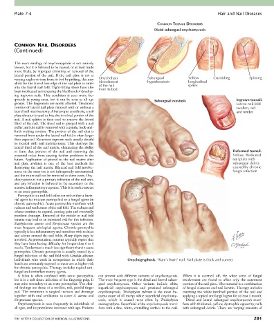
Plate 7-4                                                                                             Hair and Nail Diseases

                                                                               COMMON TOENAIL DISORDERS
                                                                              Distal subungual onychomycosis


        COMMON NAIL DISORDERS
        (Continued)


        The  exact  etiology  of  onychocryptosis  is  not  entirely
        known, but it is believed to be caused, or at least made
        more likely, by improper trimming or removal of the
        lateral  portion  of  the  nail.  If  the  nail  plate  is  cut  at
        varying angles or torn from its bed by picking, this may   Onycholysis  Subungual      Yellow       Crumbling        Splitting
        allow for the lateral free edge of the nail plate to enter   (detachment  hyperkeratosis  longitudinal
                                                                                               spikes
        into the lateral nail fold. Tight-fitting shoes have also   of the nail
                                                  from its bed)
        been implicated as increasing the likelihood of develop-
        ing  ingrown  nails.  This  condition  is  seen  more  fre-
        quently  in  young  men,  but  it  can  be  seen  in  all  age        Subungual exostosis                      Ingrown toenail.
        groups. The fingernails are rarely affected. Treatment                                                         Lateral nail fold
        consists of lateral nail plate removal with or without a                                                       swollen, red
        lateral nail matrixectomy. After proper anesthesia, a nail                                                     and tender
        plate elevator is used to free the involved portion of the
        nail. A nail splitter is then used to remove the lateral
        third of the nail. The freed nail is grasped with a nail
        puller, and the nail is removed with a gentle, back-and-
        forth rocking motion. The portion of the nail that is
        removed from under the lateral nail fold is often larger
        than expected. Recurrent ingrown nails usually should
        be  treated  with  nail  matrixectomy.  This  destroys  the
        lateral third of the nail matrix, eliminating the ability
        to  form  that  portion  of  the  nail  and  removing  the                                                   Deformed toenail.
        potential  nidus  from  causing  further  problems  in  the                                                  Yellow, thickened
        future.  Application  of  phenol  to  the  nail  matrix  after                                               nail plate with
        nail  plate  avulsion  is  one  of  the  best  methods  for                                                  subungual debris
        destroying the nail matrix. Bilateral nail fold involve-                                                     caused by chronic
        ment on the same toe is not infrequently encountered,                                                        fungal infection
        and the entire nail can be removed in these cases. Ony-
        chocryptosis is not a primary infection of the nail unit,
        and  any  infection  is  believed  to  be  secondary  to  the
        massive inflammatory response. This is in stark contrast
        to an acute paronychia.
          Paronychia is a nail fold infection with either a bacte-
        rial agent (as in acute paronychia) or a fungal agent (in
        chronic paronychia). Acute paronychia manifests with
        redness and tenderness of the nail fold. The redness and
        edema continue to expand, causing pain and eventually
        purulent drainage. Removal of the cuticle or nail fold
        trauma may lead to an increased risk for this infection.
        Staphylococcus  aureus  and  Streptococcus  species  are  the
        most  frequent  etiological  agents.  Chronic  paronychia
        typically is less inflammatory and manifests with redness
        and edema around the nail folds. Many digits may be
        involved. At presentation, patients typically report that
        they have been having difficulty for longer than 6 to 8
        weeks. Tenderness is much less significant than in acute
        paronychia. Chronic paronychia is usually caused by a
        fungal infection of the nail fold with Candida albicans.
        Individuals  who  work  in  occupations  in  which  their   Onychogryphosis. “Ram’s horn” nail. Nail plate is thick and curved.
        hands are constantly exposed to water are at higher risk
        for chronic paronychia. Therapy includes topical anti-
        fungal and antiinflammatory agents.
          A  felon  is  often  confused  with  acute  paronychia,     can present with different variants of onychomycosis.   When  it  is  curetted  off,  the  white  areas  of  fungal
        but it is a soft tissue infection of the fingertip pulp. It   The most frequent type is the distal and lateral subun-  involvement  are  found  to  affect  only  the  outermost
        may arise secondary to an acute paronychia. The clini-  gual  onychomycosis.  Other  variants  include  white   portion of the nail plate. The material is a combination
        cal findings are those of a swollen, red, painful finger   superficial  onychomycosis  and  proximal  subungual   of fungal elements and nail keratin. Therapy includes
        pad.  The  treatment  is  surgical  incision  and  drainage   onychomycosis.  Trichophyton  rubrum  is  the  most  fre-  curetting  the  white  involved  portion  of  the  nail  and
        together  with  oral  antibiotics  to  cover  S.  aureus  and   quent cause of all except white superficial onychomy-  applying a topical antifungal agent for at least 1 month.
        Streptococcus species.                    cosis,  which  is  caused  most  often  by  Trichophyton   Distal  and  lateral  subungual  onychomycosis  mani-
          Onychomycosis  is  seen  frequently  in  individuals  of   mentagrophytes. Superficial white onychomycosis mani-  fests with thickened, yellow, dystrophic appearing nails
        all ages, and its prevalence increases with age. Patients   fests with a fine, white, crumbling surface to the nail.   with subungual debris. There are varying amounts of


        THE NETTER COLLECTION OF MEDICAL ILLUSTRATIONS                                                                          201