
Page 226 - The Netter Collection of Medical Illustrations - Integumentary System_ Volume 4 ( PDFDrive )
P. 226
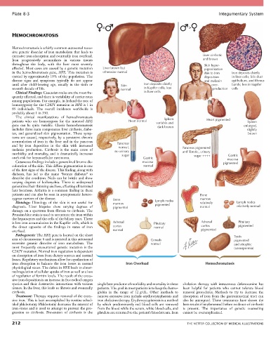
Plate 8-3                                                                                             Integumentary System


                                                   =                                          >
       HEMOCHROMATOSIS


       Hemochromatosis is a fairly common autosomal reces-
       sive genetic disorder of iron metabolism that leads to
       excessive iron absorption and eventually iron overload.                                       Liver cirrhotic
       Iron  progressively  accumulates  in  various  tissues                                        and brown
       throughout  the  body,  with  the  liver  most  severely                                      Skin hyper-
       affected. Most cases are caused by a genetic mutation   Liver brown but                       pigmentation
       in the hemochromatosis gene, HFE. This mutation is   otherwise normal                         due to iron   Iron deposits chiefly
       carried by approximately 10% of the population. The                                           deposition    in liver cells, bile duct
       disease  signs  and  symptoms  typically  do  not  appear                                     and melanin   epithelium, and fibrous
       until  after  child-bearing  age,  usually  in  the  sixth  or   Skin  Iron deposits chiefly          over-  bands; less in Kupffer
       seventh decade of life.                              normal      in Kupffer cells, less                production  cells
         Clinical Findings: Caucasian males are the most fre-           in liver cells
       quently affected, and there is variability of carrier rates
       among populations. For example, in Ireland the rate of
       homozygosity for the C282Y mutation in HFE is 1 in
       85  individuals.  The  overall  incidence  worldwide  is
       probably about 1 in 350.
         The  clinical  manifestations  of  hemochromatosis
                                                                                  Spleen
       patients  who  are  homozygous  for  the  mutated  HFE   Heart normal  variable and             Heart pigmented     Spleen
       gene  can  be  quite  variable.  Classic  hemochromatosis              dark brown                                 enlarged,
       includes three main components: liver cirrhosis, diabe-                                                            slightly
       tes,  and  generalized  skin  pigmentation.  These  symp-                                                           brown
       toms  are  caused,  respectively,  by  a  persistent  chronic
       accumulation of iron in the liver and in the pancreas
       and  by  iron  deposition  in  the  skin  with  increased   Pancreas
                                                           normal,
       melanin  production.  Cirrhosis  is  the  main  cause  of   no urinary              Pancreas pigmented
       morbidity and mortality, and it dramatically increases   sugar                      and fibrotic, urinary  Gastric
       one’s risk for hepatocellular carcinoma.                        Gastric                  sugar ++++       mucosa
         Cutaneous findings include a generalized bronze dis-          mucosa                                  pigmented
       coloration of the skin. This diffuse pigmentation is one        normal
       of the first signs of the disease. This finding, along with
       diabetes,  has  led  to  the  name  “bronze  diabetes”  to
       describe the condition. Nails can be brittle and show
       varying  degrees  of  koilonychia.  There  is  widespread
       generalized hair thinning and loss, affecting all terminal
       hair locations. Arthritis is a common finding in these
       patients and can also be seen in asymptomatic hetero-                                       Bone
       zygous carriers of the disease.                   Bone                                      marrow
         Histology:  Histology  of  the  skin  is  not  useful  for   marrow  Lymph nodes          relatively        Lymph nodes
       diagnosis.  Liver  biopsies  show  varying  degrees  of   pigmented   pigmented             normal            relatively normal
       damage on a spectrum from fibrosis to cirrhosis. The
       Prussian blue stain is used to accentuate the iron within
       the hepatocytes and the cells of the biliary tract. There
       is less iron accumulation in the Kupffer cells, which is   Adrenal    Pituitary              Adrenal           Pituitary
       the  direct  opposite  of  the  findings  in  states  of  iron   cortex  normal              cortex            pigmented
       overload.                                         normal                                     pigmented
         Pathogenesis: The HFE gene is located on the short                                                         Testis
       arm of chromosome 6 and is mutated in this autosomal                Gonads                                   pigmented
       recessive  genetic  disorder  of  iron  metabolism.  The            normal                                   and atrophic
       most  frequently  encountered  genetic  mutation  is  the                                                    (azoospermia)
       C282Y mutation. Normal iron regulation is dependent
       on absorption of iron from dietary sources and normal
       losses. Regulatory mechanisms allow for equalization of
       iron  absorption  to  balance  the  iron  losses  in  normal   Iron Overload                   Hemochromatosis
       physiological states. The defect in HFE leads to abnor-
       mal regulation of cellular uptake of iron as well as a loss
       of regulation of ferritin levels. The result of the exces-
       sive iron deposition is an increase in free radical oxygen
       species and their destructive interactions with various   single best predictor of morbidity and mortality in these   chelation  therapy  with  intravenous  deferoxamine  has
       tissues. In the liver, this leads to fibrosis and eventually   patients. The goal in most patients is to keep the hemo-  been  helpful  for  patients  who  cannot  tolerate  blood
       cirrhosis.                                globin  in  the  range  of  12 g/dL.  Other  methods  to   removal  procedures.  Methods  to  try  to  decrease  the
         Treatment: Therapy requires removal of the exces-  remove excessive iron include erythrocytapheresis and   absorption  of  iron  from  the  gastrointestinal  tract  can
       sive iron. This is best accomplished by routine sched-  iron chelation therapy. Erythrocytapheresis is a method   also  be  attempted.  These  treatments  have  shown  the
       uled phlebotomy. Phlebotomy decreases the amount of   by  which  predominantly  red  blood  cells  are  removed   best results if implemented before evidence of cirrhosis
       iron stores and is used to attempt to prevent the pro-  from the blood while the serum, white blood cells, and   is  present.  The  importance  of  genetic  counseling
       gression  to  cirrhosis.  Prevention  of  cirrhosis  is  the   platelets are returned to the patient’s bloodstream. Iron   cannot be overemphasized.

       212                                                                                   THE NETTER COLLECTION OF MEDICAL ILLUSTRATIONS