
Page 227 - The Netter Collection of Medical Illustrations - Integumentary System_ Volume 4 ( PDFDrive )
P. 227
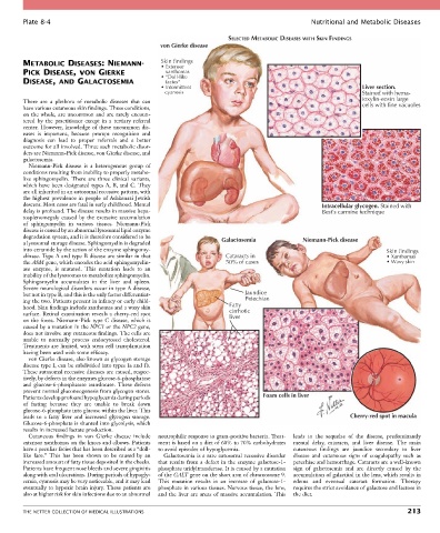
Plate 8-4                                                                                 Nutritional and Metabolic Diseases

                                                                        SELECTED METABOLIC DISEASES WITH SKIN FINDINGS
                                                   von Gierke disease

        METABOLIC DISEASES: NIEMANN-               Skin findings
                                                   • Extensor
        PICK DISEASE, vON GIERKE                    xanthomas
                                                   • “Doll-like
        DISEASE, AND GALACTOSEMIA                   facies”
                                                   • Intermittent                                                 Liver section.
                                                    cyanosis                                                      Stained with hema-
        There  are  a  plethora  of  metabolic  diseases  that  can                                               toxylin-eosin large
        have various cutaneous skin findings. These conditions,                                                   cells with fine vacuoles
        on the whole, are uncommon and are rarely encoun-
        tered  by  the  practitioner  except  in  a  tertiary  referral
        center. However, knowledge of these uncommon dis-
        eases  is  important,  because  prompt  recognition  and
        diagnosis  can  lead  to  proper  referrals  and  a  better
        outcome for all involved. Three such metabolic disor-
        ders are Niemann-Pick disease, von Gierke disease, and
        galactosemia.
          Niemann-Pick  disease  is  a  heterogenous  group  of
        conditions resulting from inability to properly metabo-
        lize  sphingomyelin.  There  are  three  clinical  variants,
        which have been designated types A, B, and C. They
        are all inherited in an autosomal recessive pattern, with
        the highest prevalence in people of Ashkenazi Jewish
        descent. Most cases are fatal in early childhood. Mental                                     Intracellular glycogen. Stained with
        delay is profound. The disease results in massive hepa-                                      Best’s carmine technique
        tosplenomegaly  caused  by  the  excessive  accumulation
        of  sphingomyelin  in  various  tissues.  Niemann-Pick
        disease is caused by an abnormal lysosomal lipid enzyme
        degradation system, and it is therefore considered to be      Galactosemia             Niemann-Pick disease
        a lysosomal storage disease. Sphingomyelin is degraded
        into ceramide by the action of the enzyme sphingomy-                                                             Skin findings
        elinase. Type A and type B disease are similar in that         Cataracts in                                      • Xanthomas
        the ASM gene, which encodes the acid sphingomyelin-            50% of cases                                      • Waxy skin
        ase  enzyme,  is  mutated.  This  mutation  leads  to  an
        inability of the lysosomes to metabolize sphingomyelin.
        Sphingomyelin  accumulates  in  the  liver  and  spleen.
        Severe neurological disorders occur in type A disease,
        but not in type B, and this is the only factor differentiat-         Jaundice
        ing the two. Patients present in infancy or early child-             Petechiae
        hood. Skin findings include xanthomas and a waxy skin           Fatty
        surface. Retinal examination reveals a cherry-red spot          cirrhotic
                                                                        liver
        on the fovea. Niemann-Pick type C disease, which is
        caused by a mutation in the NPC1 or the NPC2 gene,
        does not involve any cutaneous findings. The cells are
        unable  to  normally  process  endocytosed  cholesterol.
        Treatments are limited, with stem cell transplantation
        having been used with some efficacy.
          von Gierke disease, also known as glycogen storage
        disease type I, can be subdivided into types Ia and Ib.
        These autosomal recessive diseases are caused, respec-
        tively, by defects in the enzymes glucose-6-phosphatase
        and  glucose-6-phosphatase  translocase.  These  defects
        prevent normal gluconeogenesis from glycogen stores.
        Patients develop profound hypoglycemia during periods                      Foam cells in liver
        of  fasting  because  they  are  unable  to  break  down
        glucose-6-phosphate into glucose within the liver. This
        leads  to  a  fatty  liver  and  increased  glycogen  storage.                                        Cherry-red spot in macula
        Glucose-6-phosphate is shunted into glycolysis, which
        results in increased lactate production.
          Cutaneous  findings  in  von  Gierke  disease  include   neutrophilic response to gram-positive bacteria. Treat-  leads  to  the  sequelae  of  the  disease,  predominantly
        extensor xanthomas on the knees and elbows. Patients   ment is based on a diet of 60% to 70% carbohydrates   mental  delay,  cataracts,  and  liver  disease.  The  main
        have a peculiar facies that has been described as a “doll-  to avoid episodes of hypoglycemia.  cutaneous  findings  are  jaundice  secondary  to  liver
        like  face.”  This  has  been  shown  to  be  caused  by  an   Galactosemia is a rare autosomal recessive disorder   disease  and  cutaneous  signs  of  coagulopathy  such  as
        increased amount of fatty tissue deposited in the cheeks.   that results from a defect in the enzyme galactose-1-  petechiae and hemorrhage. Cataracts are a well-known
        Patients have frequent nose bleeds and severe gingivitis   phosphate uridyltransferase. It is caused by a mutation   sign  of  galactosemia  and  are  directly  caused  by  the
        along with oral ulcerations. During periods of hypogly-  of the GALT gene on the short arm of chromosome 9.   accumulation of galactitol in the lens, which results in
        cemia, cyanosis may be very noticeable, and it may lead   This  mutation  results  in  an  increase  of  galactose-1-  edema  and  eventual  cataract  formation.  Therapy
        eventually to hypoxic brain injury. These patients are   phosphate in various tissues. Nervous tissue, the lens,   requires the strict avoidance of galactose and lactose in
        also at higher risk for skin infections due to an abnormal   and the liver are areas of massive accumulation. This   the diet.


        THE NETTER COLLECTION OF MEDICAL ILLUSTRATIONS                                                                          213