
Page 228 - The Netter Collection of Medical Illustrations - Integumentary System_ Volume 4 ( PDFDrive )
P. 228
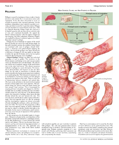
Plate 8-5                                                                                             Integumentary System

                                                                    MAIN SOURCES, CAUSES, AND SKIN FINDINGS OF PELLAGRA
       PELLAGRA
                                                            Principal sources of niacin             Principal sources of tryptophan

       Pellagra is caused by inadequate dietary intake of niacin              Whole
       (nicotinic acid, vitamin B 3 ) or its precursor amino acid,            wheat
       tryptophan.  It  has  also  been  discovered  to  occur  on            bread
       occasion  in  patients  with  carcinoid  syndrome.  In  this   Meats,                        Eggs             Milk
       syndrome, tryptophan is used entirely to produce sero-  especially liver  Whole grain cereals
       tonin, and there is none left to produce niacin. Pellagra               Principal causes of pellagra
       was first identified as a unique disease in the early 1700s
       by a Spanish physician, Gaspar Casal, who observed it                  Corn and
       in Spanish peasants who ate diets almost entirely made                 molasses           Alcohol
       of  corn  and  corn-based  foodstuffs.  He  named  the                 diet               diet
       disease “Asturian leprosy” after the region of Spain he
       was studying. An Italian physician, Francesco Frapoli,
       who studied the disease in endemic regions of northern
       Italy, later named it pellagra.                                           Deficiency of both
         Pellagra has been dominant in regions of the world                      niacin and tryptophan
       that rely heavily on corn as the main dietary staple. In
       the early twentieth century, the southern United States
       was  inundated  with  cases  of  pellagra.  Joseph  Gold-
       berger,  a  physician  and  epidemiologist  studying  the
       disease, discovered that pellagra was caused directly by
       a deficiency of vitamin B. He was unable at that time
       to isolate the specific B vitamin, but he has been given
       credit for discovering the cause of pellagra.
         Clinical Findings: Pellagra can affect any individual
       regardless  of  race  or  gender.  The  incidence  in  the
       North America and Europe is low, and cases are mainly
       caused by abnormal diets and alcoholism. The disease
       can still be seen in endemic regions of the world where
       corn  is  the  main  food  source.  The  clinical  cutaneous
       hallmark of pellagra is a severe dermatitis. The derma-
       titis  is  photosensitive,  and  exposure  to  the  sun  often
       brings  out  the  rash  or  exacerbates  it.  Patients  often
       present initially after having spent many hours outdoors
       on an early spring day. The dermatitis is symmetric and   Facial
       is manifested by eczematous patches and thin plaques   lesions;
       that tend to be tender to the touch. There is a fine line   Casal’s                                  Glove-and-stocking lesions
       of  demarcation  between  abnormal  and  normal  skin.   necklace;
       The head, neck, and arms are the most involved regions   dementia
       because of their higher level of sun exposure. The der-
       matitis  along  the  anterior  neck  and  upper  thorax  has
       been  termed  Casal  necklace.  This  is  represented  by
       weeping pink and red patches and plaques in a distribu-
       tion like that of a necklace touching the skin circumfer-
       entially around the neck. Because of its photosensitive
       nature, the dermatitis of pellagra often spares the skin
       directly  behind  the  ears  and  beneath  the  chin.  The
       nose, forehead, and cheeks are prime regions of involve-
       ment.  Non–sun-exposed  areas  can  also  be  involved,
       and  the  intertriginous  regions  are  almost  universally
       affected, including the perineum, axillae, and inframam-
       mary skin folds. The reason for the propensity to affect
       these  non–sun-exposed  regions  is  poorly  understood
       but may be related to chronic friction that induces the
       dermatitis. In the areas of involvement, small vesicula-
       tions may occur because of separation of the epidermis
       from the dermis.
         As time progresses, the dermatitis begins to desqua-
       mate. This process begins in the central portions of the
       dermatitis and spreads outward in a centrifugal manner.
       As the skin desquamates, it leaves behind red, eroded
       patches and plaques. Chronic involvement leaves per-
       manent  scarring  and  abnormal  hyperpigmentation  or   with  atrophied  papillae  are  seen  routinely  in  patients   Diarrhea is commonplace and is caused by the effect
       hypopigmentation  of  the  area.  The  epidermis  over   with  pellagra.  The  oral  and  gastrointestinal  mucous   of niacin deficiency on the gastrointestinal tract. The
       bony  prominences  (e.g.,  ulnar  head)  shows  marked   membranes may be involved. Oral ulcerations are fre-  diarrhea is watery and further complicates the patient’s
       hyperkeratosis.                           quently  seen.  Patients  routinely  complain  of  a  sore   nutritional  status  and  electrolyte  and  fluid  balances.
         Mucous  membrane  involvement  is  common  in  all   mouth and difficulty swallowing; these symptoms can   Blood and purulence may be present in the watery diar-
       vitamin deficiency states, and pellagra is no exception.   lead  to  further  lack  of  proper  nutrition,  exacerbating   rhea  as  a  result  of  ulceration  and  abscess  formation.
       Angular  cheilitis  and  a  red,  shiny,  edematous  tongue   and compounding the disease.  Ulcerations can be seen throughout the gastrointestinal

       214                                                                                   THE NETTER COLLECTION OF MEDICAL ILLUSTRATIONS