
Page 25 - The Netter Collection of Medical Illustrations - Integumentary System_ Volume 4 ( PDFDrive )
P. 25
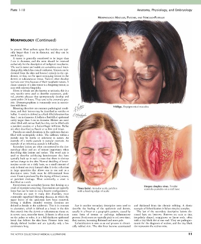
Plate 1-10                                                                            Anatomy, Physiology, and Embryology

                                                                    MORPHOLOGY: MACULES, PATCHES, AND VESICULO-PUSTULES


        MORPHOLOGY (Continued)


        be present. Most authors agree that nodules are typi-
        cally  larger  than  1 cm  in  diameter,  and  they  can  be
        much larger.
          A  tumor  is  generally  considered  to  be  larger  than
        2 cm  in  diameter,  and  the  term  should  be  reserved
        exclusively for the description of malignant neoplasms.
        The words tumor and nodule are sometimes used inter-
        changeably, which has caused confusion. Tumors can be
        elevated from the skin and located entirely in the epi-
        dermis, or they can be space-occupying lesions in the
        dermis or subcutaneous tissue. Tumors often develop
        necrosis over time because of their neoplastic nature. A
        classic example of a skin tumor is a fungating tumor, as
        seen with mycosis fungoides.
          Hives or wheals are also known as urticaria; this is a
        very  specific  term  used  to  describe  evanescent,  pink-
        red,  pruritic  plaques  that  spontaneously  develop  and
        remit within 24 hours. They tend to be extremely pru-
        ritic. Dermatographism is commonly seen in associa-
        tion with hives.                                                      Vitiligo. Depigmented macules
          Blistering disorders are common pathological condi-
        tions, and their lesions may be described as vesicles or
        bullae. A vesicle is defined as a fluid-filled elevation less
        than 1 cm in diameter. A bulla is a fluid-filled epidermal
        cavity larger than 1 cm in diameter. Blisters are most
        often filled with serous fluid, but they can be filled with
        a purulent exudate or a hemorrhagic infiltrate. Bullae
        are often described as flaccid or as firm and intact.
          Pustules are small elevations in the epidermis that are
        filled with neutrophilic debris. The infiltrate within a
        pustule  may  be  sterile  or  infectious  in  nature.  An
        example  of  a  sterile  pustule  is  pustular  psoriasis.  An
        example of an infectious pustule is folliculitis.
          Secondary lesions are often encountered in the der-
        matology  clinic  and  are  of  utmost  importance  when
        describing  skin  lesions  and  rashes.  The  word  scale  is
        used  to  describe  exfoliating  keratinocytes  that  have
        typically built up in such a mass that there is obvious
        surface change to the skin. Normal shedding of kerati-
        nocytes occurs on a daily basis, so a small amount of
        scale is found on every human’s skin. It is the collection
        in  large  quantities  that  allows  one  to  use  scale  as  a
        descriptive  term.  Scale  must  be  differentiated  from
        crust. Crust is produced by the drying of blood, serum,
        or  purulent  drainage.  Most  commonly,  a  crust  is
        described as a scab.
          Excoriations are secondary lesions that develop as a
        result of repetitive scratching. Excoriations are typically   Tinea faciei. Annular scaly patches  Herpes simplex virus. Tender
                                                                                                   vesiculo-pustules on a red base
        linear but can be seen in many bizarre configurations.  with a leading edge of scale
          Erosions  are  seen  in  many  skin  disorders,  most
        commonly superficial blistering diseases, in which the
        upper  layers  of  the  epidermis  have  been  removed,
        leaving  a  shallow,  denuded  erosion.  Erosions  are
        defined as breaks in the epidermis. This is in contrast   Scar  is  another  secondary  descriptive  term  used  to   and  thickened  from  the  chronic  rubbing.  A  classic
        to  ulceration,  which  is  defined  as  a  break  in  the  skin   describe  the  healing  of  the  epidermis  and  dermis,   example of lichenification is lichen simplex complex.
        that extends into the dermis or subcutaneous tissue or,   usually in a linear or a geographic pattern, caused by   The  last  of  the  secondary  descriptive  lesions  dis-
        in severe cases, muscular tissue. A fissure is often seen   some  form  of  trauma  or  end-stage  inflammatory   cussed  here  are  burrows.  Burrows  are  seen  as  tiny,
        on the palms or soles; it is a full-thickness epidermal   process. Fresh scars are typically pink to red; over time,   irregularly  shaped,  serpiginous  or  linear  scale,  often
        break  that  follows  the  skin  lines.  Fissures  have  very   they mature, becoming flattened and more pale.  with a tiny black dot at one end. They are pathogno-
        sharply  defined  borders  and  are  typically  only  a  few   Lichenification is seen as an end process in chroni-  monic for the diagnosis of scabies, and the tiny black
        centimeters long.                         cally rubbed skin. The skin lines become accentuated   dot represents the scabies mite.


        THE NETTER COLLECTION OF MEDICAL ILLUSTRATIONS                                                                           11