
Page 30 - The Netter Collection of Medical Illustrations - Integumentary System_ Volume 4 ( PDFDrive )
P. 30
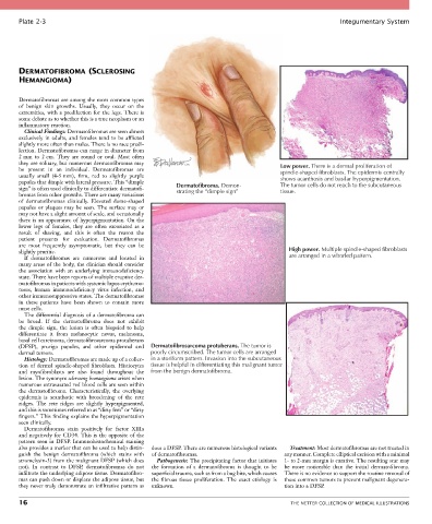
Plate 2-3                                                                                             Integumentary System


       DERMATOFIBROMA (SCLEROSING
       HEMANGIOMA)


       Dermatofibromas are among the most common types
       of  benign  skin  growths.  Usually,  they  occur  on  the
       extremities, with a predilection for the legs. There is
       some debate as to whether this is a true neoplasm or an
       inflammatory reaction.
         Clinical Findings: Dermatofibromas are seen almost
       exclusively  in  adults,  and  females  tend  to  be  afflicted
       slightly more often than males. There is no race predi-
       lection. Dermatofibromas can range in diameter from
       2 mm  to  2 cm.  They  are  round  or  oval.  Most  often
       they are solitary, but numerous dermatofibromas may                                Low power. There is a dermal proliferation of
       be  present  in  an  individual.  Dermatofibromas  are                             spindle-shaped fibroblasts. The epidermis centrally
       usually  small  (4-5 mm),  firm,  red  to  slightly  purple                        shows acanthosis and basilar hyperpigmentation.
       papules that dimple with lateral pressure. This “dimple                            The tumor cells do not reach to the subcutaneous
       sign” is often used clinically to differentiate dermatofi-  Dermatofibroma. Demon-  tissue.
                                                         strating the “dimple sign”
       bromas from other growths. There are many variations
       of  dermatofibromas  clinically.  Elevated  dome-shaped
       papules  or  plaques  may  be  seen.  The  surface  may  or
       may not have a slight amount of scale, and occasionally
       there is an appearance of hyperpigmentation. On the
       lower  legs  of  females,  they  are  often  excoriated  as  a
       result  of  shaving,  and  this  is  often  the  reason  the
       patient  presents  for  evaluation.  Dermatofibromas
       are  most  frequently  asymptomatic,  but  they  can  be
       slightly pruritic.                                                                   High power. Multiple spindle-shaped fibroblasts
         If  dermatofibromas  are  numerous  and  located  in                               are arranged in a whorled pattern.
       many areas of the body, the clinician should consider
       the  association  with  an  underlying  immunodeficiency
       state. There have been reports of multiple eruptive der-
       matofibromas in patients with systemic lupus erythema-
       tosus,  human  immunodeficiency  virus  infection,  and
       other immunosuppressive states. The dermatofibromas
       in  these  patients  have  been  shown  to  contain  more
       mast cells.
         The differential diagnosis of a dermatofibroma can
       be  broad.  If  the  dermatofibroma  does  not  exhibit
       the  dimple  sign,  the  lesion  is  often  biopsied  to  help
       differentiate  it  from  melanocytic  nevus,  melanoma,
       basal cell carcinoma, dermatofibrosarcoma protuberans
       (DFSP),  prurigo  papules,  and  other  epidermal  and  Dermatofibrosarcoma protuberans. The tumor is
       dermal tumors.                            poorly circumscribed. The tumor cells are arranged
         Histology: Dermatofibromas are made up of a collec-  in a storiform pattern. Invasion into the subcutaneous
       tion  of  dermal  spindle-shaped  fibroblasts.  Histiocytes  tissue is helpful in differentiating this malignant tumor
       and  myofibroblasts  are  also  found  throughout  the  from the benign dermatofibroma.
       lesion. The synonym sclerosing hemangioma arises when
       numerous extravasated red blood cells are seen within
       the  dermatofibroma.  Characteristically,  the  overlying
       epidermis  is  acanthotic  with  broadening  of  the  rete
       ridges.  The  rete  ridges  are  slightly  hyperpigmented,
       and this is sometimes referred to as “dirty feet” or “dirty
       fingers.” This finding explains the hyperpigmentation
       seen clinically.
         Dermatofibromas  stain  positively  for  factor  XIIIa
       and negatively for CD34. This is the opposite of the
       pattern seen in DFSP. Immunohistochemical staining
       also provides a marker that can be used to help distin-  does a DFSP. There are numerous histological variants   Treatment: Most dermatofibromas are not treated in
       guish  the  benign  dermatofibroma  (which  stains  with   of dermatofibromas.      any manner. Complete elliptical excision with a minimal
       stromelysin-3) from the malignant DFSP (which does   Pathogenesis: The precipitating factor that initiates   1- to 2-mm margin is curative. The resulting scar may
       not).  In  contrast  to  DFSP,  dermatofibromas  do  not   the  formation  of  a  dermatofibroma  is  thought  to  be   be  more  noticeable  than  the  initial  dermatofibroma.
       infiltrate the underlying adipose tissue. Dermatofibro-  superficial trauma, such as from a bug bite, which causes   There is no evidence to support the routine removal of
       mas can push down or displace the adipose tissue, but   the  fibrous  tissue  proliferation.  The  exact  etiology  is   these common tumors to prevent malignant degenera-
       they never truly demonstrate an infiltrative pattern as   unknown.                  tion into a DFSP.

       16                                                                                    THE NETTER COLLECTION OF MEDICAL ILLUSTRATIONS