
Page 29 - The Netter Collection of Medical Illustrations - Integumentary System_ Volume 4 ( PDFDrive )
P. 29
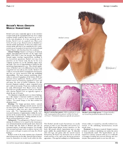
Plate 2-2                                                                                                   Benign Growths


        BECKER’S NEVUS (SMOOTH
        MUSCLE HAMARTOMA)


        Becker’s nevi most commonly appear on the shoulder
        or upper limb girdle of prepubescent boys. It is a rather
        common benign condition that is seen in up to 0.5%        Becker’s nevus
        of  the  male  population.  It  is  less  commonly  seen  in
        females.  Becker’s  nevi  are  acquired  nevi.  Most  occur
        before 10 years of age. Becker’s nevus is classified as a
        smooth muscle hamartoma. It does not contain mela-
        nocytic nevus cells and is not considered to be a mela-
        nocytic nevus. It was given its name by the dermatologist
        Samuel Becker, who first described this condition.
          Clinical Findings: Becker’s nevi begin as ill-defined,
        slightly  hyperpigmented  macules  on  the  upper  limb
        girdle.  Over  time  (1  year,  on  average),  the  hyperpig-
        mented  region  develops  hypertrichosis,  resulting  in
        its characteristic appearance. Backer’s nevi may occur
        anywhere  on  the  human  body,  but  by  far  the  most
        common  locations  are  on  the  shoulder,  upper  chest,
        and back. The area of hypertrichosis is limited to the
        underlying hyperpigmented area. The clinical signifi-
        cance of Becker’s nevi is its differentiation from large
        congenital nevi and café-au-lait macules. Becker’s nevi
        confer no increased risk for development of melanoma,
        and  they  are  rarely  associated  with  any  underlying
        abnormalities.  The  most  common  underlying  abnor-
        mality is unilateral hypoplasia of the breast, which has
        minimal  clinical  significance.  Rarely,  a  patient  with  a
        Becker’s nevus has underlying hypoplasia of bone and
        soft  tissue,  the  cause  of  which  is  unknown.  The  dif-
        ferential  diagnosis  includes  a  giant  congenital  nevus
        and a café-au-lait macule. These two conditions should
        be  easily  differentiated  from  Baker’s  nevus,  because
        they both are typically apparent at birth or soon there-
        after,  whereas  Becker’s  nevi  are  typically  acquired  at
        about the age of 10 years.
          The diagnosis is typically made on clinical findings,
        but a skin biopsy is sometime needed to confirm the
        diagnosis  if  the  nevus  is  in  an  unusual  anatomical
        location.  The  punch  biopsy  is  the  best  method  for
        obtaining tissue.
          Histology:  The  biopsy  specimen  shows  a  smooth
        muscle  hamartoma.  Multiple  smooth  muscle  fascicles
        are seen within the dermis. There is an increased ratio
        of  terminal  to  vellus  hairs  and  a  lack  of  melanocytic
        nevus  cells.  The  hyperpigmentation  results  from
        increased formation of pigmentation within the mela-  Becker’s nevus low power. Mild acanthosis is seen,  Becker’s nevus high power. Collagen bundles
        nocytes of the stratum basalis. There is no increase in   with hyperpigmentation present within the basal  surround the prominent adnexal structures.
        the number of melanocytes. Varying amounts of acan-  cell layer. Prominent sebaceous glands are present.
        thosis and hyperkeratosis are seen.
          Pathogenesis: The pathogenesis of Becker’s nevus is
        unclear. It is believed to be caused by the dermal pres-
        ence of hamartomatous smooth muscle tissue. Research   Non-Becker’s smooth muscle hamartomas are usually   release; rather, it is caused by a neurally mediated con-
        has  shown  that  the  tissue  in  Becker’s  nevi  has  an   present  at  birth  or  soon  thereafter  and  manifest  as  a   traction  of  the  underlying  hamartomatous  smooth
        increased number of androgen receptors. It is thought   small,  flesh-colored  plaque  located  anywhere  on  the   muscle tissue.
        that increased androgen levels at puberty interact with   body.  All  smooth  muscle  hamartomas  may  at  some   Treatment: No therapy is required. Surgical excision
        the excessive androgen receptors and cause the clinical   point  exhibit  the  pseudo-Darier’s  sign.  To  clinically   is likely to produce a mutilating scar unless the nevus
        findings.                                 elicit  this  sign,  one  gently  rubs  the  smooth  muscle     is  extraordinarily  small.  The  hypertrichosis  can  be
          Becker’s nevus is the most common type of smooth   hamartoma; the lesion may fasciculate due to smooth   treated for cosmetic purposes with any of a multitude
        muscle hamartoma in the skin. Smooth muscle hamar-  muscle activity, or the region may develop an urticarial   of therapies including laser removal, shaving, and elec-
        tomas by themselves are rarely found within the skin.   appearance. This sign has nothing to do with histamine   trolysis. Most patients prefer to not treat the area.


        THE NETTER COLLECTION OF MEDICAL ILLUSTRATIONS                                                                           15