
Page 31 - The Netter Collection of Medical Illustrations - Integumentary System_ Volume 4 ( PDFDrive )
P. 31
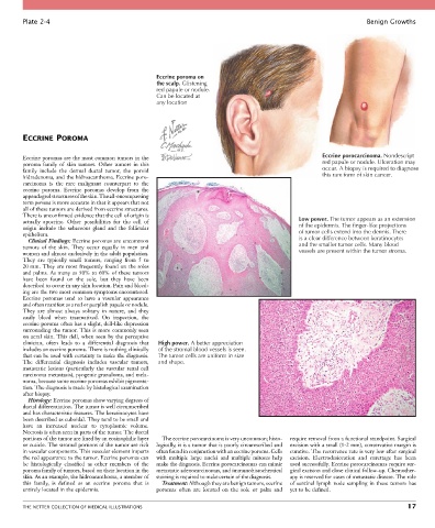
Plate 2-4                                                                                                   Benign Growths


                                                  Eccrine poroma on
                                                  the scalp. Glistening
                                                  red papule or nodule.
                                                  Can be located at
                                                  any location


        ECCRINE POROMA


        Eccrine poromas are the most common tumors in the                                             Eccrine porocarcinoma. Nondescript
        poroma  family  of  skin  tumors.  Other  tumors  in  this                                    red papule or nodule. Ulceration may
        family  include  the  dermal  ductal  tumor,  the  poroid                                     occur. A biopsy is required to diagnose
        hidradenoma, and the hidroacanthoma. Eccrine poro-                                            this rare form of skin cancer.
        carcinoma  is  the  rare  malignant  counterpart  to  the
        eccrine  poroma.  Eccrine  poromas  develop  from  the
        appendageal structures of the skin. The all-encompassing
        term poroma is more accurate in that it appears that not
        all of these tumors are derived from eccrine structures.
        There is unconfirmed evidence that the cell of origin is
        actually  apocrine.  Other  possibilities  for  the  cell  of                          Low power. The tumor appears as an extension
        origin  include  the  sebaceous  gland  and  the  follicular                           of the epidermis. The finger-like projections
        epithelium.                                                                            of tumor cells extend into the dermis. There
          Clinical Findings: Eccrine poromas are uncommon                                      is a clear difference between keratinocytes
        tumors  of  the  skin.  They  occur  equally  in  men  and                             and the smaller tumor cells. Many blood
        women and almost exclusively in the adult population.                                  vessels are present within the tumor stroma.
        They  are  typically  small  tumors,  ranging  from  5  to
        20 mm. They are most frequently found on the soles
        and palms. As many as 50% to 60% of these tumors
        have  been  found  on  the  sole,  but  they  have  been
        described to occur in any skin location. Pain and bleed-
        ing are the two most common symptoms encountered.
        Eccrine  poromas  tend  to  have  a  vascular  appearance
        and often manifest as a red or purplish papule or nodule.
        They  are  almost  always  solitary  in  nature,  and  they
        easily  bleed  when  traumatized.  On  inspection,  the
        eccrine poroma often has a slight, dell-like depression
        surrounding the tumor. This is more commonly seen
        on acral skin. This dell, when seen by the perceptive
        clinician,  often  leads  to  a  differential  diagnosis  that   High power. A better appreciation
        includes an eccrine poroma. There is nothing clinically   of the stromal blood vessels is seen.
        that can be used with certainty to make the diagnosis.   The tumor cells are uniform in size
        The  differential  diagnosis  includes  vascular  tumors,   and shape.
        metastatic  lesions  (particularly  the  vascular  renal  cell
        carcinoma metastasis), pyogenic granuloma, and mela-
        noma, because some eccrine poromas exhibit pigmenta-
        tion. The diagnosis is made by histological examination
        after biopsy.
          Histology: Eccrine poromas show varying degrees of
        ductal differentiation. The tumor is well circumscribed
        and has characteristic features. The keratinocytes have
        been described as cuboidal. They tend to be small and
        have  an  increased  nuclear  to  cytoplasmic  volume.
        Necrosis is often seen in parts of the tumor. The ductal
        portions of the tumor are lined by an eosinophilic layer   The eccrine porocarcinoma is very uncommon; histo-  require removal from a functional standpoint. Surgical
        or cuticle. The stromal portions of the tumor are rich   logically, it is a tumor that is poorly circumscribed and   excision with a small (1-2 mm), conservative margin is
        in vascular components. This vascular element imparts   often found in conjunction with an eccrine poroma. Cells   curative. The recurrence rate is very low after surgical
        the red appearance to the tumor. Eccrine poromas can   with  multiple  large  nuclei  and  multiple  mitoses  help   excision.  Electrodesiccation  and  curettage  has  been
        be  histologically  classified  as  other  members  of  the   make the diagnosis. Eccrine porocarcinomas can mimic   used successfully. Eccrine porocarcinomas require sur-
        poroma family of tumors, based on their location in the   metastatic adenocarcinomas, and immunohistochemical   gical excision and close clinical follow-up. Chemother-
        skin. As an example, the hidroacanthoma, a member of   staining is required to make certain of the diagnosis.  apy is reserved for cases of metastatic disease. The role
        this  family,  is  defined  as  an  eccrine  poroma  that  is   Treatment: Although they are benign tumors, eccrine   of sentinel lymph node sampling in these tumors has
        entirely located in the epidermis.        poromas  often  are  located  on  the  sole  or  palm  and   yet to be defined.


        THE NETTER COLLECTION OF MEDICAL ILLUSTRATIONS                                                                           17