
Page 34 - The Netter Collection of Medical Illustrations - Integumentary System_ Volume 4 ( PDFDrive )
P. 34
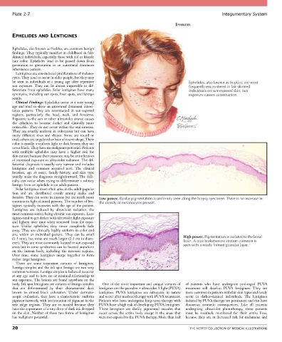
Plate 2-7                                                                                             Integumentary System

                                                                                      EPHELIDES

       EPHELIDES AND LENTIGINES


       Ephelides, also known as freckles, are common benign
       findings. They typically manifest in childhood in fair-
       skinned individuals, especially those with red or blonde
       hair  color.  Ephelides  tend  to  be  passed  down  from
       generation  to  generation  in  an  autosomal  dominant
       inheritance pattern.
         Lentigines are sun-induced proliferations of melano-
       cytes. They tend to occur in older people, but they may
       be  seen  in  individuals  at  a  young  age  after  repetitive                       Ephelides, also known as freckles, are most
       sun  exposure.  They  can  be  almost  impossible  to  dif-                           frequently encountered in fair-skinned
       ferentiate from ephelides. Solar lentigines have many                                 individuals on sun-exposed skin. Sun
       synonyms, including sun spots, liver spots, and lentigo                               exposure causes accentuation.
       senilis.
         Clinical Findings: Ephelides occur at a very young
       age and tend to show an autosomal dominant inheri-
       tance  pattern.  They  are  accentuated  in  sun-exposed
       regions,  particularly  the  head,  neck,  and  forearms.
       Exposure to the sun or other ultraviolet source causes
       the  ephelides  to  become  darker  and  clinically  more
       noticeable. They do not occur within the oral mucosa.
       They  are  usually  uniform  in  coloration  but  can  have
       many  different  sizes  and  shapes.  Some  are  round  or
       oval; others are angulated or have a bizarre shape. Their
       color is usually a uniform light to dark brown; they are
       never black. They have no malignant potential. Patients
       with  multiple  ephelides  may  have  a  higher  risk  for
       skin cancer, because their presence may be an indication
       of increased exposure to ultraviolet radiation. The dif-
       ferential diagnosis is usually very narrow and includes
       lentigines  and  common  acquired  nevi.  The  clinical
       location,  age  at  onset,  family  history,  and  skin  type
       usually make the diagnosis straightforward. The diffi-
       culty can occur when trying to differentiate a solitary
       lentigo from an ephelide in an adult patient.
         Solar lentigines most often arise in the adult popula-
       tion  and  are  distributed  evenly  among  males  and
       females. They can occur in anyone but are much more   Low power. Basilar pigmentation is uniformly seen along the biopsy specimen. There is no increase in
       common in light-skinned persons. The number of len-  the density of melanocytes present.
       tigines typically increases with the age of the patient.
       Lentigines  are  induced  by  ultraviolet  radiation,  the
       most common source being chronic sun exposure. Len-
       tigines tend to get darker with ultraviolet light exposure
       and lighten over time when removed from the expo-
       sure.  Unlike  ephelides,  they  never  completely  fade
       away. They are clinically highly uniform in color and
       size  within  an  individual  patient.  They  can  be  small
       (1-5 mm), but some are much larger (2-3 cm in diam-                                High power. Pigmentation is isolated to the basal
       eter). They are most commonly located in sun-exposed                               layer. A nice basketweave stratum corneum is
       areas but in some syndromes can be located anywhere                                seen with a nicely formed granular layer.
       on  the  human  body,  including  the  mucosal  regions.
       Over  time,  some  lentigines  merge  together  to  form
       rather large lentigines.
         There  are  some  important  variants  of  lentigines.
       Lentigo simplex and the ink spot lentigo are two very
       common versions. Lentigo simplex is believed to occur
       at any age and to have no or minimal relationship to
       sun exposure. The lesions are found anywhere on the
       body. Ink spot lentigines are variants of lentigo simplex   One of the more important and unique variants of   of  patients  who  have  undergone  prolonged  PUVA
       that  are  differentiated  by  their  characteristic  dark   lentigines are the psoralen + ultraviolet A light (PUVA)   treatment  will  develop  PUVA  lentigines.  They  are
       brown  to  almost  black  coloration.  Under  dermato-  lentigines.  PUVA  lentigines  are  iatrogenic  in  nature   more common in patients with fair skin types and rarely
       scopic  evaluation,  they  have  a  characteristic  uniform   and occur after medical therapy with PUVA treatment.   occur  in  darker-skinned  individuals.  The  lentigines
       pigment network, with accentuation of pigment in the   Patients who have undergone long-term therapy with   induced by PUVA therapy are permanent and can have
       rete  ridge  regions.  They  are  so  named  because  they   PUVA have a high risk of developing PUVA lentigines.   disastrous  cosmetic  consequences.  Like  all  patients
       have the appearance of a tiny drop of dark ink dropped   These  lentigines  are  darkly  pigmented  macules  that   undergoing  ultraviolet  phototherapy,  these  patients
       on the skin. Neither of these two forms of lentigines   occur  across  the  entire  body  except  in  the  areas  that   must  be  routinely  monitored  for  their  entire  lives,
       has malignant potential.                  were not exposed to the PUVA therapy. More than half   because  they  are  at  increased  risk  for  melanoma  and

       20                                                                                    THE NETTER COLLECTION OF MEDICAL ILLUSTRATIONS