
Page 35 - The Netter Collection of Medical Illustrations - Integumentary System_ Volume 4 ( PDFDrive )
P. 35
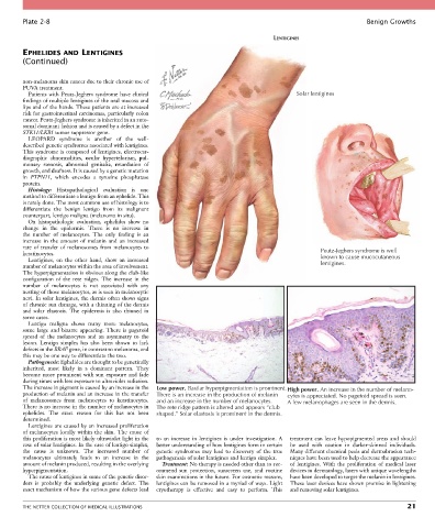
Plate 2-8                                                                                                   Benign Growths

                                                                                       LENTIGINES

        EPHELIDES AND LENTIGINES
        (Continued)


        non-melanoma skin cancer due to their chronic use of
        PUVA treatment.
          Patients with Peutz-Jeghers syndrome have clinical                                  Solar lentigines
        findings of multiple lentigines of the oral mucosa and
        lips and of the hands. These patients are at increased
        risk for gastrointestinal carcinomas, particularly colon
        cancer. Peutz-Jeghers syndrome is inherited in an auto-
        somal dominant fashion and is caused by a defect in the
        STK11/LKB1 tumor suppressor gene.
          LEOPARD  syndrome  is  another  of  the  well-
        described genetic syndromes associated with lentigines.
        This syndrome is composed of lentigines, electrocar-
        diographic  abnormalities,  ocular  hypertelorism,  pul-
        monary  stenosis,  abnormal  genitalia,  retardation  of
        growth, and deafness. It is caused by a genetic mutation
        in  PTPN11,  which  encodes  a  tyrosine  phosphatase
        protein.
          Histology:  Histopathological  evaluation  is  one
        method to differentiate a lentigo from an ephelide. This
        is rarely done. The most common use of histology is to
        differentiate  the  benign  lentigo  from  its  malignant
        counterpart, lentigo maligna (melanoma in situ).
          On  histopathologic  evaluation,  ephelides  show  no
        change  in  the  epidermis.  There  is  no  increase  in
        the  number  of  melanocytes.  The  only  finding  is  an
        increase  in  the  amount  of  melanin  and  an  increased
        rate  of  transfer  of  melanosomes  from  melanocytes  to
        keratinocytes.                                                                                Peutz-Jeghers syndrome is well
          Lentigines,  on  the  other  hand,  show  an  increased                                     known to cause mucocutaneous
        number of melanocytes within the area of involvement.                                         lentigines.
        The hyperpigmentation is obvious along the club-like
        configuration  of  the  rete  ridges.  The  increase  in  the
        number  of  melanocytes  is  not  associated  with  any
        nesting of those melanocytes, as is seen in melanocytic
        nevi. In solar lentigines, the dermis often shows signs
        of chronic sun damage, with a thinning of the dermis
        and  solar  elastosis.  The  epidermis  is  also  thinned  in
        some cases.
          Lentigo  maligna  shows  many  more  melanocytes,
        some  large  and  bizarre  appearing.  There  is  pagetoid
        spread  of  the  melanocytes  and  an  asymmetry  to  the
        lesion.  Lentigo  simplex  has  also  been  shown  to  lack
        defects in the BRAF gene, in contrast to melanoma, and
        this may be one way to differentiate the two.
          Pathogenesis: Ephelides are thought to be genetically
        inherited,  most  likely  in  a  dominant  pattern.  They
        become  more  prominent  with  sun  exposure  and  fade
        during times with less exposure to ultraviolet radiation.
        The increase in pigment is caused by an increase in the   Low power. Basilar hyperpigmentation is prominent.  High power. An increase in the number of melano-
        production of melanin and an increase in the transfer   There is an increase in the production of melanin  cytes is appreciated. No pagetoid spread is seen.
        of  melanosomes  from  melanocytes  to  keratinocytes.   and an increase in the number of melanocytes.  A few melanophages are seen in the dermis.
        There is no increase in the number of melanocytes in   The rete ridge pattern is altered and appears “club
        ephelides.  The  exact  reason  for  this  has  not  been   shaped.” Solar elastosis is prominent in the dermis.
        determined.
          Lentigines are caused by an increased proliferation
        of  melanocytes  locally  within  the  skin.  The  cause  of
        this proliferation is most likely ultraviolet light in the   to  an  increase  in  lentigines  is  under  investigation.  A   treatment  can  leave  hypopigmented  areas  and  should
        case of solar lentigines. In the case of lentigo simplex,   better understanding of how lentigines form in certain   be  used  with  caution  in  darker-skinned  individuals.
        the  cause  is  unknown.  The  increased  number  of     genetic  syndromes  may  lead  to  discovery  of  the  true   Many different chemical peels and dermabrasion tech-
        melanocytes  ultimately  leads  to  an  increase  in  the   pathogenesis of solar lentigines and lentigo simplex.  niques have been used to help decrease the appearance
        amount of melanin produced, resulting in the overlying   Treatment: No therapy is needed other than to rec-  of  lentigines.  With  the  proliferation  of  medical  laser
        hyperpigmentation.                        ommend  sun  protection,  sunscreen  use,  and  routine   devices in dermatology, lasers with unique wavelengths
          The cause of lentigines in some of the genetic disor-  skin examinations in the future. For cosmetic reasons,   have been developed to target the melanin in lentigines.
        ders  is  probably  the  underlying  genetic  defect.  The   lentigines can be removed in a myriad of ways. Light   These laser devices have shown promise in lightening
        exact mechanism of how the various gene defects lead   cryotherapy  is  effective  and  easy  to  perform.  This   and removing solar lentigines.


        THE NETTER COLLECTION OF MEDICAL ILLUSTRATIONS                                                                           21