
Page 36 - The Netter Collection of Medical Illustrations - Integumentary System_ Volume 4 ( PDFDrive )
P. 36
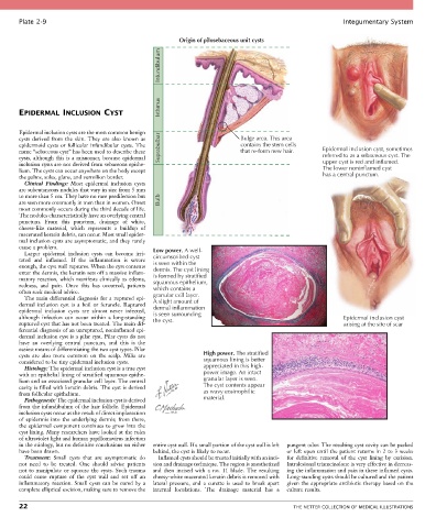
Plate 2-9                                                                                             Integumentary System

                                                         Origin of pilosebaceous unit cysts

                                                   Infundibulum


                                                   Isthmus
       EPIDERMAL INCLUSION CYST


       Epidermal inclusion cysts are the most common benign
       cysts  derived  from  the  skin.  They  are  also  known  as         Bulge area. This area
       epidermoid cysts or follicular infundibular cysts. The               contains the stem cells
       name “sebaceous cyst” has been used to describe these   Suprabulbar  that re-form new hair.    Epidermal inclusion cyst, sometimes
       cysts, although this is a misnomer, because epidermal                                          referred to as a sebaceous cyst. The
       inclusion cysts are not derived from sebaceous epithe-                                         upper cyst is red and inflamed.
       lium. The cysts can occur anywhere on the body except                                          The lower noninflamed cyst
       the palms, soles, glans, and vermilion border.                                                 has a central punctum.
         Clinical  Findings:  Most  epidermal  inclusion  cysts
       are subcutaneous nodules that vary in size from 5 mm
       to more than 5 cm. They have no race predilection but
       are seen more commonly in men than in women. Onset   Bulb
       most commonly occurs during the third decade of life.
       The nodules characteristically have an overlying central
       punctum.  From  this  punctum,  drainage  of  white,
       cheese-like  material,  which  represents  a  buildup  of
       macerated keratin debris, can occur. Most small epider-
       mal inclusion cysts are asymptomatic, and they rarely
       cause a problem.                          Low power. A well-
         Larger  epidermal  inclusion  cysts  can  become  irri-
       tated  and  inflamed.  If  the  inflammation  is  severe   circumscribed cyst
                                                 is seen within the
       enough, the cyst wall ruptures. When the cyst contents   dermis. The cyst lining
       enter the dermis, the keratin sets off a massive inflam-  is formed by stratified
       matory  reaction,  which  manifests  clinically  as  edema,   squamous epithelium,
       redness,  and  pain.  Once  this  has  occurred,  patients   which contains a
       often seek medical advice.                granular cell layer.
         The main differential diagnosis for a ruptured epi-
       dermal  inclusion  cyst  is  a  boil  or  furuncle.  Ruptured   A slight amount of
                                                 dermal inflammation
       epidermal  inclusion  cysts  are  almost  never  infected,   is seen surrounding
       although  infection  can  occur  within  a  long-standing   the cyst.                                 Epidermal inclusion cyst
       ruptured cyst that has not been treated. The main dif-                                                arising at the site of scar
       ferential diagnosis of an unruptured, noninflamed epi-
       dermal inclusion cyst is a pilar cyst. Pilar cysts do not
       have  an  overlying  central  punctum,  and  this  is  the
       easiest means of differentiating the two cyst types. Pilar
       cysts  are  also  more  common  on  the  scalp.  Milia  are   High power. The stratified
       considered to be tiny epidermal inclusion cysts.          squamous lining is better
         Histology: The epidermal inclusion cyst is a true cyst   appreciated in this high-
       with an epithelial lining of stratified squamous epithe-  power image. An intact
       lium and an associated granular cell layer. The central   granular layer is seen.
       cavity is filled with keratin debris. The cyst is derived   The cyst contents appear
       from follicular epithelium.                               as wavy eosinophilic
         Pathogenesis: The epidermal inclusion cyst is derived   material.
       from the infundibulum of the hair follicle. Epidermal
       inclusion cysts occur as the result of direct implantation
       of  epidermis  into  the  underlying  dermis;  from  there,
       the epidermal component continues to grow into the
       cyst lining. Many researchers have looked at the roles
       of ultraviolet light and human papillomavirus infection
       in the etiology, but no definitive conclusions on either   entire cyst wall. If a small portion of the cyst wall is left   pungent odor. The resulting cyst cavity can be packed
       have been drawn.                          behind, the cyst is likely to recur.      or left open until the patient returns in 2 to 3 weeks
         Treatment:  Small  cysts  that  are  asymptomatic  do   Inflamed cysts should be treated initially with an inci-  for  definitive  removal  of  the  cyst  lining  by  excision.
       not  need  to  be  treated.  One  should  advise  patients     sion and drainage technique. The region is anesthetized   Intralesional triamcinolone is very effective in decreas-
       not  to  manipulate  or  squeeze  the  cysts.  Such  trauma   and  then  incised  with  a  no.  11  blade.  The  resulting   ing the inflammation and pain in these inflamed cysts.
       could  cause  rupture  of  the  cyst  wall  and  set  off  an   cheesy-white macerated keratin debris is removed with   Long-standing cysts should be cultured and the patient
       inflammatory reaction. Small cysts can be cured by a   lateral  pressure,  and  a  curette  is  used  to  break  apart   given the appropriate antibiotic therapy based on the
       complete elliptical excision, making sure to remove the   internal  loculations.  The  drainage  material  has  a   culture results.

       22                                                                                    THE NETTER COLLECTION OF MEDICAL ILLUSTRATIONS