
Page 41 - The Netter Collection of Medical Illustrations - Integumentary System_ Volume 4 ( PDFDrive )
P. 41
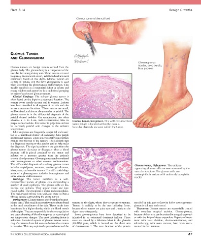
Plate 2-14                                                                                                  Benign Growths

                                                     Glomus tumor of the nail bed


        GLOMUS TUMOR
        AND GLOMANGIOMA
                                                                                               Glomangiomas
                                                                                               (tender, deep purple,
        Glomus  tumors  are  benign  tumors  derived  from  the                                blue papules)
        glomus body. The glomus body is a component of the
        vascular thermoregulatory unit. These tumors are most
        frequently encountered in early adulthood and are most
        commonly  found  on  the  digits.  Glomus  tumors  are
        solitary  in  nature,  and  the  term  glomangioma  is  used
        when describing the glomuvenous malformation. This
        usually manifests as a congenital defect in infants and
        young children and appears to be a multifocal grouping
        or mass of coalescent glomus tumors.
          Clinical  Findings:  The  solitary  glomus  tumor  is
        often found on the digit in a subungual location. The
        tumors occur equally in men and in women. Lesions
        have been described in all regions of the skin and also
        in  extracutaneous  locations.  These  tumors  are  small,
        well localized, and almost always tender or painful. The
        glomus  tumor  is  in  the  differential  diagnosis  of  the
        painful  dermal  nodules.  On  examination,  one  often
        observes  a  1-  to  2-cm,  well-circumscribed,  blue  to   Glomus tumor, low power. This well-circumscribed
        purple dermal nodule. It is tender to palpation and can   tumor lobule is located within the dermis.
        be  extremely  painful  with  changes  in  the  ambient   Vascular channels are seen within the tumor.
        temperature.
          Glomangiomas are frequently congenital and mani-
        fest  as  a  multifocal  cluster  of  coalescing,  blue-purple
        nodules and papules. There is occasionally some surface
        change over the top of the tumors. The Hildreth sign
        is a diagnostic maneuver that can be used to help make
        the diagnosis. The sign is positive if the pain from the
        glomus  tumor  decreases  or  disappears  when  a  blood
        pressure  cuff  is  placed  proximal  to  the  tumor  and
        inflated  to  a  pressure  greater  than  the  patient’s
        systolic blood pressure. Glomangiomas can be confused
        with  hemangiomas  or  other  vascular  malformations.
        The differential diagnosis of a solitary glomus tumor                                Glomus tumor, high power. The uniform-
        includes angiolipoma, neuroma, eccrine spiradenoma,                                  appearing glomus cells are seen surrounding the
        leiomyoma, and vascular tumors. The differential diag-                               vascular structure. The glomus cells are
        nosis  of  a  glomangioma  includes  hemangiomas  and                                eosinophilic in nature with uniformly basophilic
        other vascular malformations.                                                        nuclei.
          Histology:  The  tumor  manifests  as  a  well-
        circumscribed  nodule  of  glomus  cells  surrounding  a
        number of small capillaries. The glomus cells are dis-
        tinctive  and  uniform.  They  appear  round  and  have
        round nuclei. The cytoplasm is scarce and eosinophilic.
        The background stroma is myxoid, and there is often a
        fibrous capsule surrounding the entire tumor.
          Pathogenesis: Glomus tumors arise from the Sucquet-
        Hoyer canal. This canal is an arteriovenous shunt found   tumors on the digits, where they are prone to trauma.   encoded by this gene or how its defect causes gloman-
        in the small vasculature of the skin. These canals have   Trauma  is  unlikely  to  be  the  true  initiating  factor,   giomas is still not understood.
        been found in a higher density within the blood vessels   because these tumors are quite rare and trauma to the   Treatment: Glomus tumors are successfully treated
        of the digits. They are responsible for thermoregulation   digits occurs frequently.  with  complete  surgical  excision.  Glomangiomas,
        and cause shunting of blood in response to neurological   Some  glomangiomas  have  been  described  to  be   because of their size, can be excised in a staged approach
        and temperature changes. The exact initiating factor is   inherited  in  an  autosomal  dominant  fashion.  These   or with the help of tissue expanders. Reports of treat-
        unknown. Anecdotal reports of glomus tumors occur-  cases  are  caused  by  a  deletion  defect  in  the  glomulin   ment  with  laser  ablation,  electrocauterization,  and
        ring after trauma have led some to believe that trauma   (GLMN)  gene,  which  is  located  on  the  short  arm   sclerotherapy,  with  some  success,  have  been  docu-
        is causative. This may explain the preponderance of the   of  chromosome  1.  The  exact  function  of  the  protein   mented in the literature.


        THE NETTER COLLECTION OF MEDICAL ILLUSTRATIONS                                                                           27