
Page 38 - The Netter Collection of Medical Illustrations - Integumentary System_ Volume 4 ( PDFDrive )
P. 38
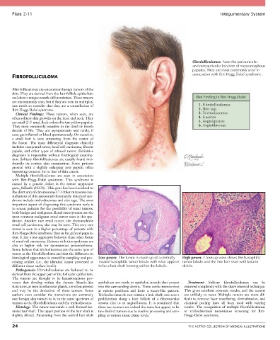
Plate 2-11                                                                                            Integumentary System


                                                                                               Fibrofolliculomas. Note the periauricular
                                                                                               and retroauricular location of monomorphous
                                                                                               papules. They are most commonly seen in
                                                                                               association with Birt-Hogg-Dubé syndrome.
       FIBROFOLLICULOMA

       Fibrofolliculomas are uncommon benign tumors of the
       skin. They are derived from the hair follicle epithelium
       and show a unique mantle differentiation. These tumors                                    Skin Finding in Birt-Hogg-Dubé
       are uncommonly seen, but if they are seen in multiples,
       one needs to consider that they are a constellation of                                     1. Fibrofolliculomas
       Birt-Hogg-Dubé syndrome.                                                                   2. Skin tags
         Clinical  Findings:  These  tumors,  when  seen,  are                                    3. Trichodiscomas
       often solitary skin growths on the head and neck. They                                     4. Lipomas
       are small (2-5 mm), flesh-colored to tan-yellow papules.                                   5. Angiolipomas
       They most commonly manifest in the third or fourth                                         6. Angiofibromas
       decade  of  life.  They  are  asymptomatic  and  rarely,  if
       ever, get inflamed or bleed spontaneously. On occasion,
       a  small  hair  is  seen  emanating  from  the  center  of
       the  lesion.  The  main  differential  diagnosis  clinically
       includes compound nevus, basal cell carcinoma, fibrous
       papule,  and  other  types  of  adnexal  tumor.  Definitive
       diagnosis  is  impossible  without  histological  examina-
       tion. Solitary fibrofolliculomas are usually found inci-
       dentally  on  routine  skin  examination.  Some  patients
       present  with  a  slightly  enlarging  new  papule,  often
       expressing concern for or fear of skin cancer.
         Multiple  fibrofolliculomas  are  seen  in  association
       with  Birt-Hogg-Dubé  syndrome.  This  syndrome  is
       caused  by  a  genetic  defect  in  the  tumor  suppressor
       gene, folliculin (FLCN) . This gene has been localized to
       the short arm of chromosome 17. Other cutaneous con-
       stellations of this autosomal dominantly inherited syn-
       drome include trichodiscomas and skin tags. The most
       important aspect of diagnosing this syndrome early is
       to  screen  patients  for  the  possibility  of  renal  tumors,
       both benign and malignant. Renal oncocytomas are the
       most common malignant renal tumor seen in this syn-
       drome.  Another  rare  renal  cancer,  the  chromophobe
       renal cell carcinoma, also may be seen. This very rare
       tumor is seen in a higher percentage of patients with
       Birt-Hogg-Dubé syndrome than in the general popula-
       tion. It has a less aggressive behavior than other forms
       of renal cell carcinoma. Patients with this syndrome are
       also  at  higher  risk  for  spontaneous  pneumothorax.
       Some believe that trichodiscomas are the same type of
       tumor as the fibrofolliculoma and that the difference in
       histological appearance is caused by sampling and pro-  Low power. The tumor is made up of a centrally  High power. Close-up view shows the basophilic
       cessing  artifact  (i.e.,  the  identical  tumor  processed  at   located basophilic tumor lobule with what appears tumor lobule and the fine hair shaft with keratin
       different tissue surface levels).         to be a hair shaft forming within the lobule.  debris.
         Pathogenesis:  Fibrofolliculomas  are  believed  to  be
       derived from the upper part of the follicular epithelium.
       The  tumors  are  thought  to  be  hamartomatous  pro-
       cesses  that  develop  within  the  dermis.  Mantle-like   epithelium are cords or epithelial strands that project   Treatment:  Solitary  fibrofolliculomas  can  be
       structures, as seen in sebaceous glands, are often present   into the surrounding dermis. These cords interconnect   removed completely with the shave removal technique.
       and  may  be  the  derivation  of  these  tumors.  Some   at  various  positions  and  form  a  weave-like  pattern.   This  gives  excellent  cosmetic  results,  and  the  tumors
       authors  even  consider  the  manteloma  (an  extremely   Trichodiscomas do not contain a hair shaft; one sees a   are  unlikely  to  recur.  Multiple  tumors  are  more  dif-
       rare benign skin tumor) to be in the same spectrum of   proliferation  along  a  hair  follicle  of  a  fibrovascular   ficult to remove; laser resurfacing, dermabrasion, and
       tumors as the fibrofolliculoma and the trichodiscoma.  stroma  akin  to  an  angiofibroma.  It  is  postulated  that   chemical  peeling  have  all  been  used  with  varying
         Histology: The tumor surrounds a well-formed ter-  these two tumors are indeed the same but appear to be   results.  The  recognition  of  multiple  fibrofolliculomas
       minal hair shaft. The upper portion of the hair shaft is   two distinct tumors due to routine processing and sam-  or  trichodiscomas  necessitates  screening  for  Birt-
       slightly dilated. Emanating from the central hair shaft   pling at various tissue plane levels.  Hogg-Dubé  syndrome.

       24                                                                                    THE NETTER COLLECTION OF MEDICAL ILLUSTRATIONS