
Page 39 - The Netter Collection of Medical Illustrations - Integumentary System_ Volume 4 ( PDFDrive )
P. 39
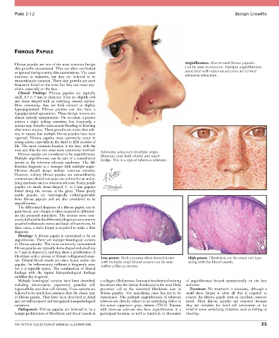
Plate 2-12                                                                                                  Benign Growths


        FIBROUS PAPULE


        Fibrous papules are one of the most common benign                                   Angiofibromas. Also termed fibrous papules.
        skin growths encountered. They are often overlooked                                 Can be seen in isolation. Multiple angiofibromas
        or ignored during routine skin examinations. The exact                              associated with tuberous sclerosis are termed
        incidence  is  unknown,  but  they  are  believed  to  be                           adenoma sebaceum.
        extraordinarily common. These skin growths are most
        frequently found on the nose, but they can occur any-
        where, especially on the face.
          Clinical  Findings:  Fibrous  papules  are  typically
        small, 0.5 to 5 mm in diameter. They are slightly oval
        and  dome  shaped  with  an  overlying  smooth  surface.
        Most  commonly,  they  are  flesh  colored  to  slightly
        hyperpigmented.  Fibrous  papules  can  also  have  a
        hypopigmented appearance. These benign tumors are
        almost  entirely  asymptomatic.  On  occasion,  a  patient
        notices  a  slight  itching  sensation;  less  frequently,  a
        patient may describe spontaneous bleeding or bleeding
        after minor trauma. These growths are most often soli-
        tary in nature, but multiple fibrous papules have been
        reported.  Fibrous  papules  most  commonly  occur  in
        young adults, especially in the third to fifth decades of
        life. The most common location is the face, with the
        nose and chin the two areas most commonly involved.  Adenoma sebaceum (multiple angio-
          Fibrous papules are considered to be angiofibromas.
        Multiple angiofibromas can be part of a constellation   fibromas) over both cheeks and nasal
                                                  bridge. This is a sign of tuberous sclerosis.
        known  as  the  tuberous  sclerosis  syndrome.  The  dif-
        ferential diagnosis in a teenager with multiple angio-
        fibromas  should  always  include  tuberous  sclerosis.
        However,  solitary  fibrous  papules  are  extraordinarily
        common and should not cause one to look for an under-
        lying syndrome such as tuberous sclerosis. Pearly penile
        papules  are  small,  dome-shaped,  1-  to  2-mm  papules
        found  along  the  corona  of  the  glans.  These  pearly
        penile  papules  are  histologically  indistinguishable
        from  fibrous  papules  and  are  also  considered  to  be
        angiofibromas.
          The differential diagnosis of a fibrous papule can be
        quite broad, and a biopsy is often required to differenti-
        ate  the  potential  mimickers.  The  entities  most  com-
        monly included in the differential diagnosis are common
        acquired melanocytic nevus and basal cell carcinoma. In
        these cases, a shave biopsy is required to make a firm
        diagnosis.
          Histology:  A  fibrous  papule  is  considered  to  be  an
        angiofibroma. There are multiple histological variants
        of fibrous papules. The most commonly encountered
        fibrous papules are typically dome shaped and small (up
        to 5 mm in diameter), and they show a proliferation of
        fibroblasts with a stroma of fibrotic collagenized mate-  Low power. Well-circumscribed dermal tumor  High power. Fibroblasts are the main cell type
        rial. Dilated blood vessels are often found within the   with multiple small blood vessels can be seen  along with the blood vessels.
        papules. An inflammatory infiltrate is frequently seen,   within a fibrous stroma.
        but it is typically sparse. The combination of clinical
        findings  with  the  typical  histopathological  findings
        solidifies the diagnosis.
          Multiple  histological  variants  have  been  described,   a collagen-filled stroma. Immunohistochemical staining   of  angiofibromas  located  symmetrically  on  the  face
        including  pleomorphic,  pigmented,  granular  cell,   has shown that the dermal dendrocyte is the most likely   and nose.
        hypercellular, and clear cell variants. These variants are   precursor  cell  to  the  abnormal  fibroblasts  seen  in   Treatment:  No  treatment  is  necessary,  although  a
        believed to be much less common than the classic type   fibrous  papules.  The  underlying  cause  has  yet  to  be   small  shave  biopsy  is  often  all  that  is  required  to
        of fibrous papule. They have been described in detail   determined.  The  multiple  angiofibromas  of  tuberous   remove the fibrous papule with an excellent cosmetic
        and are well accepted and recognized histopathological   sclerosis are directly related to an underlying defect in   result.  Most  fibrous  papules  are  removed  because
        variations.                               the  tumor  suppressor  gene,  tuburin  (TSC2).  Patients   they  are  mistaken  for  basal  cell  carcinomas  or  for
          Pathogenesis:  Fibrous  papules  are  believed  to  be  a   with  tuberous  sclerosis  also  have  angiofibromas  in  a   relief of some underlying irritation, such as itching or
        benign proliferation of fibroblasts and blood vessels in   periungual location, as well as hundreds to thousands   bleeding.


        THE NETTER COLLECTION OF MEDICAL ILLUSTRATIONS                                                                           25