
Page 37 - The Netter Collection of Medical Illustrations - Integumentary System_ Volume 4 ( PDFDrive )
P. 37
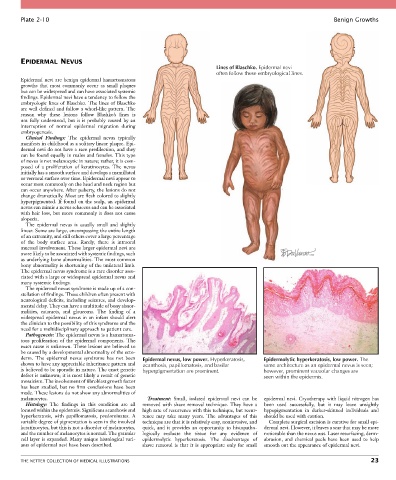
Plate 2-10                                                                                                  Benign Growths


        EPIDERMAL NEVUS
                                                                            Lines of Blaschko. Epidermal nevi
                                                                            often follow these embryological lines.
        Epidermal  nevi  are  benign  epidermal  hamartomatous
        growths  that  most  commonly  occur  as  small  plaques
        but can be widespread and can have associated systemic
        findings. Epidermal nevi have a tendency to follow the
        embryologic lines of Blaschko. The lines of Blaschko
        are well defined and follow a whorl-like pattern. The
        reason  why  these  lesions  follow  Blashko’s  lines  is
        not  fully  understood,  but  it  is  probably  caused  by  an
        interruption  of  normal  epidermal  migration  during
        embryogenesis.
          Clinical  Findings:  The  epidermal  nevus  typically
        manifests in childhood as a solitary linear plaque. Epi-
        dermal nevi do not have a race predilection, and they
        can be found equally in males and females. This type
        of nevus is not melanocytic in nature; rather, it is com-
        posed  of  a  proliferation  of  keratinocytes.  The  nevus
        initially has a smooth surface and develops a mamillated
        or verrucal surface over time. Epidermal nevi appear to
        occur most commonly on the head and neck region but
        can occur anywhere. After puberty, the lesions do not
        change dramatically. Most are flesh colored to slightly
        hyperpigmented.  If  found  on  the  scalp,  an  epidermal
        nevus can mimic a nevus sebaceus and can be associated
        with hair loss, but more commonly it does not cause
        alopecia.
          The  epidermal  nevus  is  usually  small  and  slightly
        linear. Some are large, encompassing the entire length
        of an extremity, and still others cover a large percentage
        of  the  body  surface  area.  Rarely,  there  is  intraoral
        mucosal involvement. These larger epidermal nevi are
        more likely to be associated with systemic findings, such
        as underlying bone abnormalities. The most common
        bony abnormality is shortening of the unilateral limb.
        The epidermal nevus syndrome is a rare disorder asso-
        ciated with a large or widespread epidermal nevus and
        many systemic findings.
          The epidermal nevus syndrome is made up of a con-
        stellation of findings. These children often present with
        neurological deficits, including seizures, and develop-
        mental delay. They can have a multitude of bony abnor-
        malities,  cataracts,  and  glaucoma.  The  finding  of  a
        widespread  epidermal  nevus  in  an  infant  should  alert
        the clinician to the possibility of this syndrome and the
        need for a multidisciplinary approach to patient care.
          Pathogenesis: The epidermal nevus is a hamartoma-
        tous  proliferation  of  the  epidermal  components.  The
        exact cause is unknown. These lesions are believed to
        be caused by a developmental abnormality of the ecto-
        derm.  The  epidermal  nevus  syndrome  has  not  been   Epidermal nevus, low power. Hyperkeratosis,  Epidermolytic hyperkeratosis, low power. The
        shown to have any appreciable inheritance pattern and   acanthosis, papillomatosis, and basilar  same architecture as an epidermal nevus is seen;
        is believed to be sporadic in nature. The exact genetic   hyperpigmentation are prominent.  however, prominent vacuolar changes are
        defect is unknown; it is most likely a result of genetic                            seen within the epidermis.
        mosaicism. The involvement of fibroblast growth factor
        has  been  studied,  but  no  firm  conclusions  have  been
        made. These lesions do not show any abnormalities of
        melanocytes.                                Treatment:  Small,  isolated  epidermal  nevi  can  be   epi dermal nevi. Cryotherapy with liquid nitrogen has
          Histology:  The  findings  in  this  condition  are  all   removed  with  shave  removal  technique.  They  have  a   been  used  successfully,  but  it  may  leave  unsightly
        located within the epidermis. Significant acanthosis and   high rate of recurrence with this technique, but recur-  hypopigmentation  in  darker-skinned  individuals  and
        hyperkeratosis,  with  papillomatosis,  predominates.  A   rence  may  take  many  years.  The  advantages  of  this   should be used with caution.
        variable degree of pigmentation is seen in the involved   technique are that it is relatively easy, noninvasive, and   Complete surgical excision is curative for small epi-
        keratinocytes, but this is not a disorder of melanocytes,   quick,  and  it  provides  an  opportunity  to  histopatho-  dermal nevi. However, it leaves a scar that may be more
        and the number of melanocytes is normal. The granular   logically  evaluate  the  tissue  for  any  evidence  of     noticeable than the nevus was. Laser resurfacing, derm-
        cell layer is expanded. Many unique histological vari-  epi dermolytic  hyperkeratosis.  The  disadvantage  of   abrasion,  and  chemical  peels  have  been  used  to  help
        ants of epidermal nevi have been described.  shave  removal  is  that  it  is  appropriate  only  for  small     smooth out the appearance of epidermal nevi.


        THE NETTER COLLECTION OF MEDICAL ILLUSTRATIONS                                                                           23