
Page 44 - The Netter Collection of Medical Illustrations - Integumentary System_ Volume 4 ( PDFDrive )
P. 44
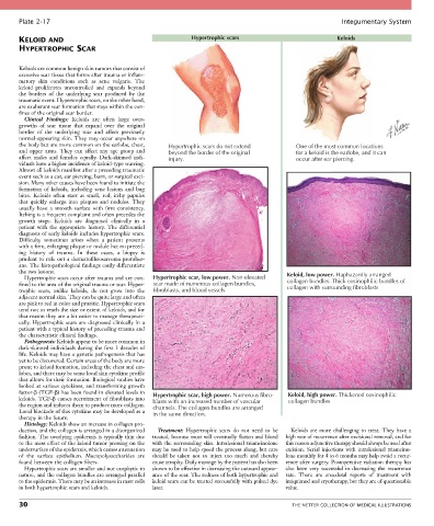
Plate 2-17                                                                                            Integumentary System

       KELOID AND                                            Hypertrophic scars                            Keloids
       HYPERTROPHIC SCAR


       Keloids are common benign skin tumors that consist of
       excessive scar tissue that forms after trauma or inflam-
       matory  skin  conditions  such  as  acne  vulgaris.  The
       keloid  proliferates  uncontrolled  and  expands  beyond
       the  borders  of  the  underlying  scar  produced  by  the
       traumatic event. Hypertrophic scars, on the other hand,
       are exuberant scar formation that stays within the con-
       fines of the original scar border.
         Clinical  Findings:  Keloids  are  often  large  over-
       growths  of  scar  tissue  that  expand  over  the  original
       border  of  the  underlying  scar  and  affect  previously
       normal-appearing skin. They may occur anywhere on
       the body but are more common on the earlobe, chest,   Hypertrophic scars do not extend  One of the most common locations
       and  upper  arms.  They  can  affect  any  age  group  and   beyond the border of the original  for a keloid is the earlobe, and it can
       affect  males  and  females  equally.  Dark-skinned  indi-  injury.                    occur after ear piercing.
       viduals have a higher incidence of keloid-type scarring.
       Almost all keloids manifest after a preceding traumatic
       event such as a cut, ear piercing, burn, or surgical exci-
       sion. Many other causes have been found to initiate the
       formation  of  keloids,  including  acne  lesions  and  bug
       bites.  Keloids  often  start  as  small,  red,  itchy  papules
       that  quickly  enlarge  into  plaques  and  nodules.  They
       usually  have  a  smooth  surface  with  firm  consistency.
       Itching is a frequent complaint and often precedes the
       growth  stage.  Keloids  are  diagnosed  clinically  in  a
       patient  with  the  appropriate  history.  The  differential
       diagnosis of early keloids includes hypertrophic scars.
       Difficulty  sometimes  arises  when  a  patient  presents
       with a firm, enlarging plaque or nodule but no preced-
       ing  history  of  trauma.  In  these  cases,  a  biopsy  is
       prudent to rule out a dermatofibrosarcoma protuber-
       ans. The histopathological findings easily differentiate
       the two lesions.                                                                    Keloid, low power. Haphazardly arranged
         Hypertrophic scars occur after trauma and are con-  Hypertrophic scar, low power. Non-elevated
       fined to the area of the original trauma or scar. Hyper-  scar made of numerous collagen bundles,  collagen bundles. Thick eosinophilic bundles of
                                                                                           collagen with surrounding fibroblasts
       trophic  scars,  unlike  keloids,  do  not  grow  into  the   fibroblasts, and blood vessels
       adjacent normal skin. They can be quite large and often
       are pink to red in color and pruritic. Hypertrophic scars
       tend not to reach the size or extent of keloids, and for
       that reason they are a bit easier to manage therapeuti-
       cally. Hypertrophic scars are diagnosed clinically in a
       patient with a typical history of preceding trauma and
       the characteristic clinical findings.
         Pathogenesis: Keloids appear to be more common in
       dark-skinned individuals during the first 3 decades of
       life. Keloids may have a genetic pathogenesis that has
       yet to be discovered. Certain areas of the body are more
       prone to keloid formation, including the chest and ear-
       lobes, and there may be some local skin cytokine profile
       that allows for their formation. Biological studies have
       looked at various cytokines, and transforming growth
       factor-β (TGF-β) has been found in elevated levels in   Hypertrophic scar, high power. Numerous fibro-  Keloid, high power. Thickened eosinophilic
       keloids. TGF-β causes recruitment of fibroblasts into   blasts with an increased number of vascular  collagen bundles
       the region and induces them to produce more collagen.   channels. The collagen bundles are arranged
       Local blockade of this cytokine may be developed as a   in the same direction.
       therapy in the future.
         Histology: Keloids show an increase in collagen pro-
       duction, and the collagen is arranged in a disorganized   Treatment:  Hypertrophic  scars  do  not  need  to  be   Keloids  are  more  challenging  to  treat.  They  have  a
       fashion. The overlying epidermis is typically thin due   treated, because most will eventually flatten and blend   high rate of recurrence after excisional removal, and for
       to the mass effect of the keloid tumor pressing on the   with  the  surrounding  skin.  Intralesional  triamcinolone   this reason adjunctive therapy should always be used after
       undersurface of the epidermis, which causes attenuation   may be used to help speed the process along, but care   excision.  Serial  injections  with  intralesional  triamcino-
       of  the  surface  epithelium.  Mucopolysaccharides  are   should  be  taken  not  to  inject  too  much  and  thereby     lone monthly for 4 to 6 months may help avoid a recur-
       found between the collagen fibers.        cause atrophy. Daily massage by the patient has also been   rence after surgery. Postoperative radiation therapy has
         Hypertrophic scars are smaller and not exophytic in   shown to be effective in decreasing the outward appear-  also  been  very  successful  in  decreasing  the  recurrence
       nature, and the collagen bundles are arranged parallel   ance of the scar. The redness of both hypertrophic and   rate.  There  are  anecdotal  reports  of  treatment  with
       to the epidermis. There may be an increase in mast cells   keloid scars can be treated successfully with pulsed dye   imiquimod and cryotherapy, but they are of questionable
       in both hypertrophic scars and keloids.   laser.                                    value.

       30                                                                                    THE NETTER COLLECTION OF MEDICAL ILLUSTRATIONS