
Page 42 - The Netter Collection of Medical Illustrations - Integumentary System_ Volume 4 ( PDFDrive )
P. 42
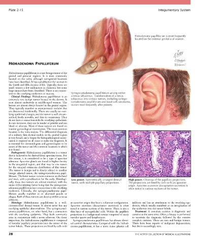
Plate 2-15                                                                                            Integumentary System


                                                                                             Hidradenoma papilliferum is most frequently
                                                                                             located on the external genitalia of women.


       HIDRADENOMA PAPILLIFERUM


       Hidradenoma papilliferum is a rare benign tumor of the
       genital  and  perianal  regions.  It  is  most  commonly
       located  on  the  vulva,  although  extragenital  locations
       have been described. It has a predilection for women in
       the fourth and fifth decades of life. Typically, these are
       small tumors a few millimeters in diameter, but some
       large tumors have been described. There is no connec-
       tion to the overlying epidermis or mucosa.  Syringocystadenoma papilliferum arising within
         Clinical  Findings:  Hidradenoma  papilliferum  is  an   a nevus sebaceous. Transformation of a nevus
       extremely rare benign tumor located in the dermis. It   sebaceous into various tumors, including syringo-
       seen  almost  exclusively  in  middle-aged  women.  The   cystadenoma papilliferum and basal cell carcinoma,
       lesions are almost always located in the genital region.   occurs most frequently after puberty.
       They typically manifest as asymptomatic nodules that
       are discovered incidentally. There are usually no over-
       lying epidermal changes, and the tumor is well circum-
       scribed, freely movable, and firm in consistency. They
       do not have a connection with the overlying epithelium.
       In rare instances, they can be tender or pruritic and can
       bleed or ulcerate. Most of these tumors are found on
       routine gynecological examination. The most common
       location is the labia majora. The differential diagnosis
       of a solitary, firm dermal nodule in the genital region
       is very broad, and a biopsy for histopathological exami-
       nation is required in all cases to make the diagnosis. It
       is essential for dermatologists and gynecologists to be
       aware of this tumor and the common locations in which
       it is found.
         Pathogenesis: Hidradenoma papilliferum is a tumor
       that is believed to be derived from apocrine tissue. For
       this  reason,  it  is  considered  to  be  a  type  of  apocrine
       adenoma. Apocrine glands are found in higher density
       in the anogenital region, and that may be one reason
       for  the  unequal  cutaneous  distribution  of  this  tumor.
       The tumor is benign and is closely related to another
       benign adnexal tumor, the syringocystadenoma papil-
       liferum. The latter tumor is more common on the head
       and neck, with a predilection for the scalp. Histologi-  Low power. Symmetrically arranged dermal  High power. Close-up of the papillary projections.
       cally, these two tumors are almost identical, with the   tumor, with multiple papillary projections.  The projections are lined by cells with an apocrine
       major differentiating factor being that the syringocyst-                         origin. Apocrine secretion (decapitation secretion) is
       adenoma papilliferum has a connection to the overlying                           often noted in various sections of the tumor.
       epidermis.  Clinically,  the  syringocystadenoma  papil-
       liferum  usually  manifest  as  an  ulcerated  papule  or
       plaque. Both of these tumors can develop within a nevus
       sebaceus.
         Histology:  Hidradenoma  papilliferum  is  a  well-  an apocrine origin that have a columnar configuration.   infiltrate and has an attachment to the overlying epi-
       circumscribed  dermal  tumor.  It  almost  never  has  any   Apocrine  secretion  (decapitation  secretion)  is  often   dermis, which usually manifests as an invagination of
       overlying epithelial abnormalities. The syringocystad-  noted in various sections of the tumor. There is also a   the epidermis into the tumor lobule.
       enoma papilliferum, on other hand, has a connection   thin layer of myoepithelial cells. Within the papillary   Treatment:  A  complete  excision  is  diagnostic  and
       with  the  overlying  epidermis.  They  both  commonly   projections is a background stroma composed of many   curative at the same time. Often, a biopsy is performed
       arise in conjunction with a nevus sebaceus. On closer   vascular spaces and lymphocytes.  to  ascertain  the  diagnosis,  followed  by  the  curative
       inspection, the hidradenoma papilliferum is composed   Syringocystadenoma papilliferum has almost identi-  complete excision. These are rare and benign tumors.
       of vascular papillary projections into the center of the   cal central characteristics. Compared with the hidrad-  There  have  been  reports  of  malignant  degeneration,
       tumor lobule. These projections are lined by cells with   enoma  papilliferum,  it  has  a  more  dense  plasma  cell   but this is exceedingly rare.

       28                                                                                    THE NETTER COLLECTION OF MEDICAL ILLUSTRATIONS