
Page 47 - The Netter Collection of Medical Illustrations - Integumentary System_ Volume 4 ( PDFDrive )
P. 47
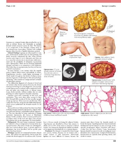
Plate 2-20                                                                                                  Benign Growths


                                                                     Massive
                                                                     lipoma of       Sectioned lipoma composed
                                                                     axilla          of yellow fat lobules with narrow
        LIPOMA                                                                       intervening fibrous septa


        Lipomas are common benign skin growths that can be
        seen  as  solitary  lesions  and  frequently  as  multiple
        dermal  nodules  scattered  about  the  skin.  The  lipoma
        is  an  overgrowth  of  the  fibrofatty  adipose  tissue  in
        the  subcutaneous  tissue  plane.  Patients  with  multiple
        lipomas often describe a familial inheritance pattern.
          Clinical Findings: Lipomas are often small (1-2 cm),
        soft, subcutaneous nodules that are slow growing and                    Large liposarcoma
        freely  moveable  underneath  the  skin.  Some  lipomas                 of posterior thigh          Lipoma. Soft, rubbery, freely
        become quite large (>5 cm in diameter), and they can                                                moveable dermal nodule
        be a cause for concern due to interference with move-
        ment  and  the  possibility  of  malignant  degeneration
        into a liposarcoma. There are no overlying epidermal
        changes, and there is no connection to the epidermis.
        Most often they are asymptomatic, but they can become
        painful if traumatized.                    Liposarcoma. CT scan
          In  stark  contrast,  a  rare  variant  called  the  angioli-  reveals mixture of benign
        poma is almost always tender and multiple in nature.   (low-density) and sar-
        Angiolipomas  contain  a  much  higher  percentage  of   comatous (high-density)
        blood vessels throughout the lobule of adipose tissue,   areas of tumor.
        and the diagnosis is made based on this histopathologi-
        cal finding. These tumors are benign and have no famil-                                          Liposarcoma. Excised tumor with
        ial inheritance pattern.                                                                         muscle at margin; tumor darker
          The differential diagnosis of a lipoma is broad and                                            and firmer than benign lipoma
        can include other dermal tumors; however, the clinical
        examination findings are often diagnostic. Occasionally,
        a small lipoma can be confused with an epidermal inclu-
        sion  cyst,  pilar  cyst,  lymph  node,  or  adnexal  tumor.
        Large,  freely  movable,  rubbery  nodules  that  are  slow
        growing are easily diagnosed clinically as lipomas.
          Lipomas  occur  most  commonly  on  the  trunk  and
        extremities.  They  most  often  affect  women  in  their
        third through fifth decades of life but can affect people
        of any age and sex. There is no race predilection. They
        rarely affect the face, except for the subfrontalis lipoma,
        which  occurs  underneath  the  frontalis  muscle  on  the
        forehead.
          Rare syndromes of adipose tissue have been described,
        including benign symmetric lipomatosis, adiposis dolo-
        rosa (Dercum’s disease), and familial multiple lipomato-
        sis.  The  best  described  of  these  syndromes  is  benign   Low power. Adipocytes with varying amounts  High power. Mature adipocytes are the main
        symmetric  lipomatosis,  also  known  as  Madelung’s   of fibrous tissue and blood vessels  component of the tumor.
        disease. In this condition, there is massive proliferation
        of adipose tissue on the neck and upper arms of men.
        The patients take on the appearance of a body builder.
          Pathogenesis: The exact cause is unknown. Lipomas   have  a  fibrous  capsule  enclosing  the  adipose  lobules.   surgeon  must  dissect  below  the  frontalis  muscle  to
        are believed to be an overgrowth of normal tissue in a   Angiolipomas  are  described  as  those  fatty  tumors  in   locate the lipoma. Small lipomas have been treated with
        normal location. The tumor lobules are indistinguish-  which 10% to 50% of the mass is composed of blood   intralesional steroid injection to take advantage of the
        able from normal adipose tissue. A genetic pattern of   vessels. The various rare lipomatosis variants are identi-  steroid’s  atrophogenic  effects.  Injections  with  deoxy-
        inheritance  has  been  described,  but  no  specific  gene   cal in appearance histologically to a common lipoma.  cholate  have  also  been  effective.  Large,  fast-growing
        defect has been located.                    Treatment: No therapy is required for these benign   lipomas should be removed to rule out malignant trans-
          Histology: Lipomas are composed of mature adipose   skin  tumors.  Solitary  lipomas  can  be  treated  with  a   formation into a liposarcoma. Compared with lipomas,
        tissue. The lobules are separated by fibrous septa that   simple  excision  or  with  liposuction.  Subfrontalis   liposarcomas  are  typically  faster  growing,  firmer,  and
        contain the blood supply for the adipose cells. Lipomas   lipomas  are  more  difficult  to  remove,  because  the   tender in nature.


        THE NETTER COLLECTION OF MEDICAL ILLUSTRATIONS                                                                           33