
Page 48 - The Netter Collection of Medical Illustrations - Integumentary System_ Volume 4 ( PDFDrive )
P. 48
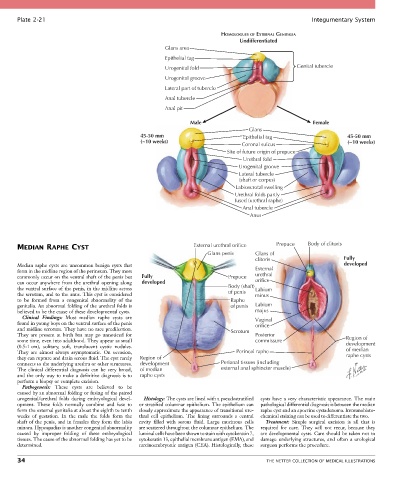
Plate 2-21                                                                                            Integumentary System

                                                                             HOMOLOGUES OF EXTERNAL GENITALIA
                                                                                    Undifferentiated
                                                          Glans area
                                                          Epithelial tag
                                                          Urogenital fold                              Genital tubercle
                                                          Urogenital groove
                                                          Lateral part of tubercle
                                                          Anal tubercle

                                                          Anal pit

                                                                   Male                                      Female
                                                                                       Glans
                                                  45-50 mm                           Epithelial tag                      45-50 mm
                                                  (~10 weeks)                                                            (~10 weeks)
                                                                                     Coronal sulcus
                                                                               Site of future origin of prepuce
                                                                                     Urethral fold
                                                                                   Urogenital groove
                                                                                    Lateral tubercle
                                                                                    (shaft or corpus)
                                                                                  Labioscrotal swelling
                                                                                  Urethral folds partly
                                                                                  fused (urethral raphe)
                                                                                     Anal tubercle
                                                                                       Anus


       MEDIAN RAPHE CYST                                            External urethral orifice   Prepuce    Body of clitoris
                                                                         Glans penis     Glans of
                                                                                         clitoris                       Fully
       Median  raphe  cysts  are  uncommon  benign  cysts  that                                                         developed
       form in the midline region of the perineum. They most                             External
       commonly occur on the ventral shaft of the penis but   Fully             Prepuce  urethral
       can occur anywhere from the urethral opening along   developed                    orifice
       the ventral surface of the penis, in the midline across                  Body (shaft) Labium
       the scrotum, and to the anus. This cyst is considered                    of penis  minus
       to  be  formed  from  a  congenital  abnormality  of  the                 Raphe
       genitalia. An abnormal folding of the urethral folds is                   of penis  Labium
       believed to be the cause of these developmental cysts.                            majus
         Clinical  Findings:  Most  median  raphe  cysts  are                            Vaginal
       found in young boys on the ventral surface of the penis                           orifice
       and midline scrotum. They have no race predilection.                      Scrotum
       They  are  present  at  birth  but  may  go  unnoticed  for                       Posterior
       some time, even into adulthood. They appear as small                              commissure                      Region of
       (0.5-1 cm),  solitary,  soft,  translucent  cystic  nodules.                                                      development
       They  are  almost  always  asymptomatic.  On  occasion,                     Perineal raphe                        of median
       they can rupture and drain serous fluid. The cyst rarely   Region of                                              raphe cysts
       connects to the underlying urethra or other structures.   development  Perianal tissues (including
       The  clinical  differential  diagnosis  can  be  very  broad,   of median  external anal sphincter muscle)
       and the only way to make a definitive diagnosis is to   raphe cysts
       perform a biopsy or complete excision.
         Pathogenesis:  These  cysts  are  believed  to  be
       caused by an abnormal folding or fusing of the paired
       urogenital/urethral  folds  during  embryological  devel-  Histology: The cysts are lined with a pseudostratified   cysts have a very characteristic appearance. The main
       opment.  These  folds  normally  combine  and  fuse  to   or stratified columnar epithelium. The epithelium can   pathological differential diagnosis is between the median
       form the external genitalia at about the eighth to tenth   closely approximate the appearance of transitional ure-  raphe cyst and an apocrine cystadenoma. Immunohisto-
       weeks  of  gestation.  In  the  male  the  folds  form  the     thral  cell  epithelium.  The  lining  surrounds  a  central   chemical staining can be used to differentiate the two.
       shaft of the penis, and in females they form the labia   cavity  filled  with  serous  fluid.  Large  mucinous  cells     Treatment:  Simple  surgical  excision  is  all  that  is
       minora. Hypospadias is another congenital abnormality   are scattered throughout the columnar epithelium. The   required  for  cure.  They  will  not  recur,  because  they
       caused  by  improper  folding  of  these  embryological   luminal cells have been shown to stain with cytokeratin 7,   are developmental cysts. Care should be taken not to
       tissues. The cause of the abnormal folding has yet to be   cytokeratin 13, epithelial membrane antigen (EMA), and   damage  underlying  structures,  and  often  a  urological
       determined.                               carcinoembryonic antigen (CEA). Histologically, these   surgeon performs the procedure.

       34                                                                                    THE NETTER COLLECTION OF MEDICAL ILLUSTRATIONS