
Page 49 - The Netter Collection of Medical Illustrations - Integumentary System_ Volume 4 ( PDFDrive )
P. 49
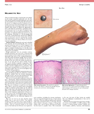
Plate 2-22                                                                                                  Benign Growths

                                                                                       BLUE NEVI


        MELANOCYTIC NEVI


        There are numerous types of melanocytic nevi, includ-
        ing  the  benign  congenital  melanocytic  nevi,  the  blue           Blue nevus
        nevi, and the common acquired melanocytic nevi. Atyp-
        ical and dysplastic nevi are discussed with melanoma in
        the section on malignant growths. Evaluation of mela-
        nocytic nevi is one of the dermatologist’s most common
        and important tasks. Every patient who enters a derma-
        tologist’s  office  should  be  offered  the  opportunity  to
        have a full-body skin examination, specifically evaluat-
        ing melanocytic nevi for any signs of malignant trans-
        formation and or de novo melanoma production. The
        importance of evaluating melanocytic nevi is to screen
        for  melanoma.  Melanoma  is  a  life-threatening  skin
        cancer that, if discovered early, can be cured. Different
        types of melanocytic nevi have varying rates of malig-
        nant transformation, and it is critical for the clinician
        to be aware of those nevi that are likely to be encoun-
        tered on a daily basis.
          Clinical Findings: Melanocytic nevi can be classified
        both  clinically  and  histopathologically.  The  common
        acquired melanocytic nevus is a clinical diagnosis, and
        if the lesion is biopsied, it may show some evidence of
        atypia or dysplasia of melanocytes. It is for this reason
        that a universally accepted classification of melanocytic
        nevi has yet to be adopted.
          Benign  melanocytic  nevi  are  extremely  common.
        Virtually all humans have some form of these growths
        on their body. Common acquired melanocytic nevi are
        universally found and can have varying morphologies.
        They affect males and females equally. They are uncom-
        mon  at  birth  but  increase  in  number  over  the  first  4
        decades of life, after which the number typically stabi-
        lizes.  As  one  ages,  the  nevi  tend  to  slowly  involute.
        They can be macular or papular in appearance. Most
        are uniform and symmetric in size and color. They can
        be flesh colored or slightly brown in coloration. They
        tend to grow proportionally as a child grows or as an
        adult gains weight. They also can become slightly larger
        and darker during pregnancy.
          There is a risk for malignant degeneration into mela-
        noma, and changes in color, size, symmetry, or border
        should  be  assessed.  Nevi  that  become  symptomatic,
        especially  pruritic,  and  nevi  that  spontaneously  bleed
        should be evaluated and biopsied appropriately.
          Blue nevi are unique benign melanocytic tumors that
        have  a  characteristic  clinical  and  histological  pattern.
        These nevi tend to be small, to be located on the dorsal
        aspect of the hands or feet, and to have a bluish to blue-
        gray coloration due to their location within the dermis.
        The blue color is believed to result from the Tyndall   Blue nevus low power. The epidermis appears  Blue nevus high power. Elongated pigmented
        effect. This is a process by which various wavelengths   normal. The dermis is filled with spindle-shaped  melanocytes are appreciated with multiple dermal
        of  light  are  absorbed  preferentially,  and  the  reflected   melanocytes and many melanophages.  melanophages. The melanocytes are interspersed
        light or color that is seen depends on the material and                             between the collagen bundles.
        depth  of  the  substance  being  illuminated.  Blue  nevi
        share similar histological characteristics with the nevus
        of Ota, nevus of Ito, and Mongolian spots. However,
        the clinical appearance is so different that these lesions
        are  not  considered  in  the  differential  diagnosis  of  a
        blue nevus.                               occur  anywhere,  including  the  mucous  membranes.   is  the  case,  but  most  of  these  lesions  are  actually
          Blue  nevi  can  occur  at  any  age,  and  they  appear   They are frequently biopsied because of their unusual   blue  nevi.  Malignant  transformation  of  blue  nevi  is
        equally  often  in  men  and  in  women.  They  typically   coloration. They are small and usually can be removed   extremely rare.
        manifest as small (2-5 mm), oval or round macules or   easily with a punch biopsy that is 1 mm larger than the   Multiple blue nevi can be seen in the Carney complex,
        papules. They are well circumscribed with nice, distinct   lesion.  Patients  often  give  a  history  of  having  been   also known as the NAME or LAMB syndrome. This
        borders.  They  are  commonly  located  on  the  dorsal   stabbed  with  a  pencil  during  childhood  and  believe     complex  of  clinical  findings  includes  multiple  blue
        aspect of the hands and feet but have been reported to   that  the  lesion  is  a  graphite  tattoo.  This  occasionally     nevi,  lentigines,  ephelides,  myxomas,  atrial  myxomas,


        THE NETTER COLLECTION OF MEDICAL ILLUSTRATIONS                                                                           35