
Page 50 - The Netter Collection of Medical Illustrations - Integumentary System_ Volume 4 ( PDFDrive )
P. 50
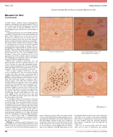
Plate 2-23                                                                                            Integumentary System

                                                               COMMON ACQUIRED NEVI AND GIANT CONGENITAL MELANOCYTIC NEVI


       MELANOCYTIC NEVI
       (Continued)


       testicular  tumors,  pituitary  tumors,  psammomatous
       melanotic schwannomas, and adrenal tumors. This is a
       rare syndrome that has been determined to be caused
       by  a  genetic  defect  in  the  gene  PRKAR1A.  This  is  a
       tumor suppressor gene that encodes a protein kinase A
       subunit.
         Congenital melanocytic nevi can be divided clinically
       into distinct subtypes based on size (small, medium, and
       giant).  Small  congenital  nevi  are  the  most  common
       type; they are defined as those nevi smaller than 2 cm
       in greatest diameter. These nevi occur with equal fre-
       quency in males and females and have no race predilec-
       tion. Some authors estimate their prevalence at about
       1% of the population. These nevi are typically described
       as well-defined macules, papules, or plaques. They are
       hyperpigmented compared with the normal surround-
       ing skin. They are almost always uniform in color and
       symmetric. Over time, some 50% develop terminal hair   Common acquired nevus                Atypical/dysplastic nevus with
       growth within  the  nevi. The  risk  of  malignant  trans-                                  surrounding solar lentigines
       formation  in  these  small  congenital  nevi  is  low  and
       approaches that of the common acquired melanocytic
       nevi. Melanoma can arise in these nevi at any point in
       the patient’s life but usually after puberty.
         Medium-sized  congenital  melanocytic  nevi  are
       defined  as  those  that  have  a  diameter  between  2  and
       20 cm. They have the same risk of malignant transfor-
       mation as small congenital nevi. They occur equally in
       males and females and can be seen in about 1% of the
       population. They can occur anywhere on the body.
         Giant  or  large  congenital  melanocytic  nevi,  also
       known as “bathing trunk” nevi, are important clinically
       in  many  ways.  First,  they  have  an  increased  risk  of
       malignant transformation. This transformation can be
       difficult to discern clinically until the lesions are quite
       large. Most melanomas develop in a dermal or subcu-
       taneous  location,  which  make  them  difficult  to  assess
       clinically.  Melanomas  typically  occur  before  puberty,
       and they have been reported to occur in as many as 15%
       of giant congenital nevi. The risk of malignant trans-
       formation is higher in axial nevi than in acral nevi. For
       this reason, these lesions are treated more aggressively,
       and  patients  with  large  congenital  melanocytic  nevi
       need lifelong, frequent routine follow-up. These nevi
       occur  equally  in  men  and  women  and  in  any  racial
       group. They affect the truncal region more often than
       any other region of the body.                            Nevus spilus                              Halo nevus
         The significant finding of neurocutaneous melanosis
       occurs at a higher rate in patients with large congenital
       nevi of the trunk. These nevi almost always occur over
       the majority of the trunk, and they can have any number
       of satellite melanocytic nevi. Patients with large truncal
       congenital melanocytic nevi should undergo magnetic
       resonance  imaging  (MRI)  of  the  nervous  system  to
       evaluate  for  neurocutaneous  melanosis.  Patients  with
       neurocutaneous  melanosis  are  at  a  high  risk  (almost
       50%)  for  development  of  leptomeningeal  melanoma,
       which  is  almost  always  fatal.  A  multidisciplinary   normal melanocytes found within the stratum basalis.   histologically. Based on the location of the melanocyte
       approach to care for these patients is required, includ-  They are round and uniform in shape and show increas-  nests, they can be classified as junctional, intraepider-
       ing  the  patient’s  pediatrician,  dermatologist,  neurolo-  ing maturation with depth in the dermis. Maturation of   mal, dermal, or compound nevi. A junctional nevus has
       gist, and neurosurgeon.                   nevi cells implies a decrease in the ratio of nuclear to   its  nests  arranged  along  the  basement  membrane
         Histology:  In  common  acquired  melanocytic  nevi,   cytoplasmic volume and an overall decrease in the size   zone,  whereas  a  compound  nevus  has  epidermal  and
       the melanocytes are arranged symmetrically in a lateral   of the melanocytes. The melanocytes are still uniform   dermal nests.
       fashion. They are arranged in nests. The nested mela-  in size and shape at various depths within the dermis;   Blue  nevi  are  located  entirely  within  the  dermis.
       nocytes do not have the typical dendritic appearance of   they are not symmetric vertically. Many forms are seen   These  nevi  are  made  of  melanocytes  that  resemble

       36                                                                                    THE NETTER COLLECTION OF MEDICAL ILLUSTRATIONS