
Page 51 - The Netter Collection of Medical Illustrations - Integumentary System_ Volume 4 ( PDFDrive )
P. 51
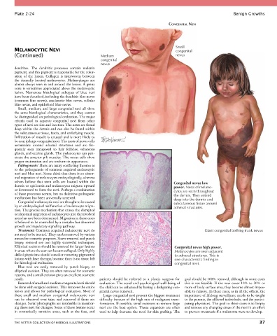
Plate 2-24                                                                                                  Benign Growths

                                                                                    CONGENITAL NEVI


                                                                                        Small
        MELANOCYTIC NEVI                                                                congenital
        (Continued)                                Medium                               nevus
                                                   congenital
                                                   nevus
        dendrites.  The  dendritic  processes  contain  melanin
        pigment, and this pigment is responsible for the color-
        ation  of  the  lesion.  Collagen  is  interwoven  between
        the  dermally  located  melanocytes.  Melanophages  are
        almost always seen in and around the lesion. A grenz
        zone is sometimes appreciated above the melanocytic
        lesion.  Numerous  histological  subtypes  of  blue  nevi
        have been described, including the dendritic blue nevus
        (common blue nevus), amelanotic blue nevus, cellular
        blue nevus, and epithelioid blue nevus.
          Small,  medium,  and  large  congenital  nevi  all  show
        the  same  histological  characteristics,  and  they  cannot
        be distinguished on pathological evaluation. The major
        criteria  used  to  separate  congenital  nevi  from  other
        types of nevi are size and location. The nests are found
        deep within the dermis and can also be found within
        the subcutaneous tissue, fascia, and underlying muscle.
        Infiltration of muscle is unusual and is more likely to
        be seen in large congenital nevi. The nests of nevus cells
        accumulate  around  adnexal  structures  and  are  fre-
        quently  seen  juxtaposed  to  hair  follicles,  sebaceous
        glands, and eccrine glands. The melanocytes can pen-
        etrate the arrector pili muscles. The nevus cells show
        proper maturation and are uniform in appearance.
          Pathogenesis: There are many conflicting theories as
        to the pathogenesis of common acquired melanocytic
        nevi and blue nevi. Some think that there is an abnor-
        mal migration of melanocytes embryologically, whereas
        others  believe  that  stem  cells  are  located  within  the                  Congenital nevus low
        dermis or epidermis and melanocytes migrate upward                             power. Nests of melano-
        or downward to form the nevi. Perhaps a combination                            cytes are seen throughout
        of these processes occurs, but no definitive pathogenic                        the dermis. They extend
        mechanism has been universally accepted.                                       deep into the dermis and
          Congenital melanocytic nevi are thought to be caused                         subcutaneous tissue around
        by an embryological malfunction of melanocyte migra-                           adnexal structures.
        tion. The precise mechanism that causes the disrupted
        or abnormal migration of melanocytes into the involved
        areas has not been determined. Migration in these cases
        is believed to be controlled by a complex but abnormal
        growth and regulatory signaling pathway.
          Treatment: Common acquired melanocytic nevi do                                              Giant congenital bathing trunk nevus
        not need to be treated. They can be removed by various
        means for cosmetic purposes. Shave removal and punch
        biopsy  removal  are  two  highly  successful  techniques.
        Elliptical excision should be reserved for larger lesions                      Congenital nevus high power.
        in areas where the scar can be camouflaged. Only highly                        Melanocytes are seen adjacent
        skilled physicians should consider removing pigmented                          to adnexal structures. This is
        lesions with laser therapy, because there is no tissue left                    one characteristic finding in
        for histological evaluation.                                                   congenital nevi.
          Blue  nevi  are  easily  removed  by  punch  biopsy  or
        elliptical excision. They are often removed for cosmetic
        reasons, and a small excision gives an excellent cosmetic
        result.                                   patients  should  be  referred  to  a  plastic  surgeon  for   goal should be 100% removal, although in some cases
          Removal of small and medium congenital nevi should   evaluation. The social and psychological well-being of   this is not feasible. If the nevi cover 10% to 30% or
        be done with surgical excision. This removes the entire   the child can be enhanced by having a disfiguring con-  more of body surface area, they become almost impos-
        lesion and allows for pathological evaluation. Most of   genital nevus removed.     sible to remove. In these cases, as in all the others, the
        these  small  and  medium  congenital  melanocytic  nevi   Large congenital nevi present the biggest treatment   importance of lifelong surveillance needs to be taught
        can  be  observed  over  time  and  removed  if  there  are   difficulty because of the high rate of malignant trans-  to the parents, the afflicted individuals, and the partici-
        changes. Serial photographs are invaluable in monitor-  formation. If possible, serial excisions to remove large   pating physicians. The goal in these cases is to biopsy
        ing these nevi for changes. Some of these lesions occur   nevi  are  the  best  option.  Tissue  expanders  are  often   and remove any changing areas of the nevi in an effort
        in  cosmetically  sensitive  areas,  such  as  the  face,  and   used to help decrease the need for skin grafting. The   to prevent metastasis if a melanoma were to develop.


        THE NETTER COLLECTION OF MEDICAL ILLUSTRATIONS                                                                           37