
Page 52 - The Netter Collection of Medical Illustrations - Integumentary System_ Volume 4 ( PDFDrive )
P. 52
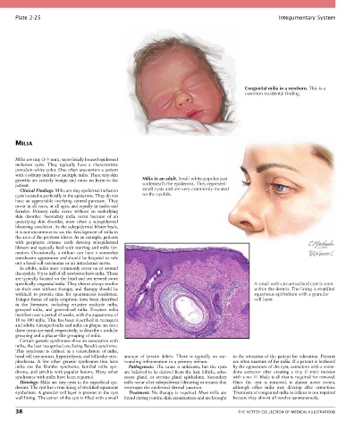
Plate 2-25                                                                                            Integumentary System


                                                                                                Congenital milia in a newborn. This is a
                                                                                                common incidental finding.


       MILIA


       Milia are tiny (1-3 mm), superficially located epidermal
       inclusion  cysts.  They  typically  have  a  characteristic
       porcelain-white color. One often encounters a patient
       with a solitary milium or multiple milia. These tiny skin
       growths are entirely benign and cause no harm to the   Milia in an adult. Small white papules just
       patient.                                         underneath the epidermis. They represent
         Clinical Findings: Milia are tiny epidermal inclusion   small cysts and are very commonly located
       cysts located superficially in the epidermis. They do not   on the eyelids.
       have an appreciable overlying central punctum. They
       occur in all races, at all ages, and equally in males and
       females.  Primary  milia  occur  without  an  underlying
       skin  disorder.  Secondary  milia  occur  because  of  an
       underlying  skin  disorder,  most  often  a  subepidermal
       blistering condition. As the subepidermal blister heals,
       it is not uncommon to see the development of milia in
       the area of the previous blister. As an example, patients
       with  porphyria  cutanea  tarda  develop  subepidermal
       blisters and typically heal with scarring and milia for-
       mation.  Occasionally,  a  milium  can  have  a  somewhat
       translucent appearance and should be biopsied to rule
       out a basal cell carcinoma or an intradermal nevus.
         In adults, milia most commonly occur on or around
       the eyelids. Up to half of all newborns have milia. These
       are typically located on the head and are termed more
       specifically congenital milia. They almost always resolve                                   A small well-circumscribed cyst is seen
       on their own without therapy, and therapy should be                                         within the dermis. The lining is stratified
       withheld  to  provide  time  for  spontaneous  resolution.                                  squamous epithelium with a granular
       Unique forms of milia eruptions have been described                                         cell layer.
       in  the  literature,  including  eruptive  multiple  milia,
       grouped  milia,  and  generalized  milia.  Eruptive  milia
       manifest over a period of weeks, with the appearance of
       10 to 100 milia. This has been described in teenagers
       and adults. Grouped milia and milia en plaque are rare;
       these terms are used, respectively, to describe a nodular
       grouping and a plaque-like grouping of milia.
         Certain genetic syndromes show an association with
       milia, the best recognized one being Bazek’s syndrome.
       This  syndrome  is  defined  as  a  constellation  of  milia,
       basal cell carcinomas, hypotrichosis, and follicular atro-  amount  of  keratin  debris.  There  is  typically  no  sur-  to the attention of the patient for education. Patients
       phoderma.  A  few  other  genetic  syndromes  that  have   rounding inflammation in a primary milium.  are often unaware of the milia. If a patient is bothered
       milia  are  the  Rombo  syndrome,  familial  milia  syn-  Pathogenesis:  The  cause  is  unknown,  but  the  cysts   by the appearance of the cyst, extraction with a come-
       drome, and atrichia with papular lesions. Many other   are believed to be derived from the hair follicle, seba-  done  extractor  after  creating  a  tiny  (1-mm)  incision
       syndromes with milia have been reported.  ceous  gland,  or  eccrine  gland  epithelium.  Secondary   with a no. 11 blade is all that is required for removal.
         Histology: Milia are tiny cysts in the superficial epi-  milia occur after subepidermal blistering or trauma that   Once  the  cyst  is  removed,  it  almost  never  recurs,
       dermis. The cyst has a true lining of stratified squamous   interrupts the epidermal-dermal junction.  although  other  milia  may  develop  after  extraction.
       epithelium. A granular cell layer is present in the cyst   Treatment: No therapy is required. Most milia are   Treatment of congenital milia in infants is not required
       wall lining. The center of the cyst is filled with a small   found during routine skin examinations and are brought   because they almost all resolve spontaneously.

       38                                                                                    THE NETTER COLLECTION OF MEDICAL ILLUSTRATIONS