
Page 53 - The Netter Collection of Medical Illustrations - Integumentary System_ Volume 4 ( PDFDrive )
P. 53
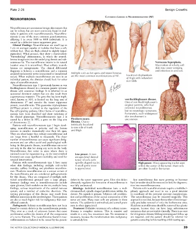
Plate 2-26                                                                                                  Benign Growths

                                                                          CUTANEOUS LESIONS IN NEUROFIBROMATOSIS (NF)
        NEUROFIBROMA


        Neurofibromas are uncommon benign skin tumors that
        can be solitary but are more commonly found in mul-
        tiples in patients with neurofibromatosis. Neurofibro-
        matosis is one of the more common genodermatoses,
        afflicting  1  in  every  3000  to  4000  individuals.  It  is
        caused by a defective tumor suppressor gene.
          Clinical  Findings:  Neurofibromas  are  small  (up  to
        1 cm on average) papules or nodules that have a soft,
        rubbery feel. They are flesh colored to slightly hyper-
        pigmented. When pressed, they show a characteristic
        “buttonholing”  phenomenon,  in  which  the  neurofi-
        broma invaginates into the underlying dermis and sub-
        cutaneous fat. The neurofibroma returns to its natural                                               Verrucous hyperplasia.
        location once it is unconfined. Most solitary neurofi-                                               Maceration of velvety-soft
        bromas  are  asymptomatic.  The  clinical  differential                                              skin may cause weeping
        diagnosis  is  between  a  neurofibroma  and  a  common                                              and infection in crevices.
        acquired melanocytic nevus (compound or intradermal   Multiple café-au-lait spots and neurofibromas  Localized elephantiasis
        nevus).  When  multiple  neurofibromas  are  seen  in  an   are the most common manifestations of NF.  of thigh with redundant
        individual patient, the clinician should look for other                          skin folds
        signs of neurofibromatosis.
          Neurofibromatosis type 1 (previously known as von
        Recklinghausen disease) is a common genetic systemic
        disease  with  cutaneous  findings.  It  is  inherited  in  an
        autosomal  dominant  pattern  but  can  also  result  from
        a spontaneous mutation. The gene that has been impli-
        cated,  known  as  NF1,  is  located  on  the  long  arm  of                    von Recklinghausen disease.
        chromosome  17  and  encodes  the  tumor  suppressor                            One of von Recklinghausen’s
        protein, neuro fibromin. This guanosine triphosphatase                          original patients, who had
        (GTPase)  protein  is  critical  in  the  regulation  of  the                   extensive neurofibromas
        Ras cell signaling pathway. Other forms of neurofibro-                          but no neurologic symptoms.
        matosis  have  been  described  and  show  variations  of                       Fortunately, such widespread
        the  clinical  phenotype.  Neurofibromatosis  type  2  is                       skin involvement is
        caused  by  a  defect  in  NF2,  a  gene  on  the  long  arm   Plexiform neuro-  uncommon.
        of  chromosome  22.                        fibroma. Charac-
          Patients with neurofibromatosis type 1 begin devel-  teristically localized
        oping  neurofibromas  at  puberty,  and  the  lesions   to one side of trunk
        increase  in  number  dramatically  over  their  life  span.   and thigh
        They are often larger than solitary neurofibromas and
        can  range  from  a  handful  to  thousands.  The  sheer
        number of neurofibromas can cause significant disfig-
        urement and can affect social and psychological well-
        being. In this genetic disease, neurofibromas can occur
        not only in the skin but along any nerve in the body.
        Neurofibromas  that  occur  in  areas  where  there  is
        minimal room for expansion (e.g., in the intervertebral   Low power. A non-
        foramen) can cause significant morbidity and need for   encapsulated dermal
        surgical  intervention.                      tumor of cells with
          Patients  with  neurofibromatosis  type  1  have  many   spindle-shaped nuclei.        High power. Wavy-appearing nuclei seen
        other  skin  findings,  including  multiple  café-au-lait   A small grenz zone           within the center of the tumor. Mast cells
        macules,  axillary  freckling,  and  plexiform  neurofibro-  is appreciated.             are often found in the tumor.
        mas. Plexiform neurofibromas are a unique variant of
        the  neurofibroma  and  are  considered  pathognomonic
        for this disease. They are composed of multiple indi-
        vidual  neurofibromas  grouped  into  a  large  plaque.     defect in the tumor suppressor gene. How this defect   Any  neurofibroma  that  starts  growing  or  becomes
        Systemic  findings  seen  in  neurofibromatosis  include   ultimately regulates the formation of neurofibromas is   hard or tender should be removed to look for degenera-
        optic gliomas, Lisch nodules on the iris, multiple bony   not fully understood.     tion into neurofibrosarcoma.
        findings,  various  impairments  of  the  central  nervous   Histology:  Individual  neurofibromas  have  a  well-  Patients with neurofibromatosis require a multidisci-
        system,  and  a  number  of  endocrine  disorders.  The   circumscribed, spindle-shaped proliferation within the   plinary  approach  and  need  to  see  a  good  internist
        varying  phenotypes  of  this  disease  may  result  from     dermis. No capsule is present. Schwann cell prolifera-  to coordinate all the potential systemic complications.
        different mutations in the involved gene. These patients   tion and proliferation of the axonal components of the   The  neurofibromas  may  be  removed  surgically.  This
        are also at much higher risk for malignancy than non-  nerve  are  seen.  Many  mast  cells  are  present  in  these   approach is not ideal, because the number of lesions typi-
        afflicted controls.                       tumors. The epidermis is uninvolved, and a small grenz   cally precludes removal of only the bothersome ones.
          Pathogenesis: Solitary neurofibromas have not been   zone is often appreciated.   Plexiform neurofibromas should be removed by a plastic
        found to contain defects in the neurofibromin protein.   Treatment: Definitive treatment of a solitary neuro-  surgeon,  because  they  can  have  large  subcutaneous
        They  arise  as  a  result  of  unknown  factors  that  cause   fibroma  is  complete  excision.  This  is  curative  and   extensions that are not visible clinically. There is no cure
        proliferation within the dermis of all the components   results in a very low recurrence rate. No treatment is   for this genetic disease; lifelong screening and follow-up
        of a nerve filament. The neurofibromas found in neu-  necessary, because the transformation into malignancy   are  required,  and  the  patient  should  be  referred  for
        rofibromatosis are believed to be caused by the genetic   is extremely low.         genetic counseling before reaching child-bearing age.


        THE NETTER COLLECTION OF MEDICAL ILLUSTRATIONS                                                                           39