
Page 74 - The Netter Collection of Medical Illustrations - Integumentary System_ Volume 4 ( PDFDrive )
P. 74
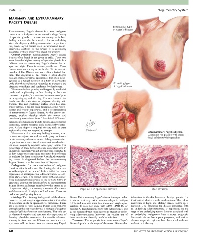
Plate 3-9                                                                                             Integumentary System

       MAMMARY AND EXTRAMAMMARY
       PAGET’S DISEASE
                                                                               Eczematous type
                                                                               of Paget’s disease
       Extramammary  Paget’s  disease  is  a  rare  malignant
       tumor that typically occurs in areas with a high density
       of  apocrine  glands.  It  is  most  commonly  an  isolated
       finding  but  can  also  be  a  marker  for  an  underlying
       visceral malignancy of the gastrointestinal or genitouri-
       nary tract. Paget’s disease is an intraepidermal adeno-
       carcinoma  confined  to  the  breast;  it  is  commonly
       associated with an underlying breast malignancy.
         Clinical  Findings:  Extramammary  Paget’s  disease
       is most often found in the groin or axilla. These two
       areas have the highest density of apocrine glands. It is
       believed  that  extramammary  Paget’s  disease  has  an
       apocrine origin. There is no race predilection. These
       tumors  most  commonly  occur  in  the  fifth  to  seventh
       decades of  life.  Women are  more  often  affected  than
       men.  The  diagnosis  of  this  tumor  is  often  delayed
       because of its eczematous appearance. It is often misdi-
       agnosed as a fungal infection or a form of dermatitis.
       Only after the area has not responded to therapy is the                Ulcerating type
       diagnosis considered and confirmed by skin biopsy.                     of Paget’s disease
         The tumor is slow growing and is typically a red-pink
       patch  with  a  glistening  surface.  Itching  is  the  most
       common complaint, but patients also complain of pain,
       burning, stinging, and bleeding. The area is sore to the
       touch,  and  there  are  areas  of  pinpoint  bleeding  with
       friction.  The  red,  glistening  surface  often  has  small
       white patches. This has been described as the “straw-
       berries and cream” appearance, and it is characteristic
       of  extramammary  Paget’s  disease.  As  the  cancer  pro-
       gresses,  erosions  develop  within  the  tumor,  and
       occasionally ulcerations form. The clinical differential
       diagnosis is often among Paget’s disease, an eczematous
       dermatitis, inverse psoriasis, and a dermatophyte infec-
       tion.  A  skin  biopsy  is  required  for  any  rash  in  these
       regions that does not respond to therapy.                                                      Extramammary Paget’s disease.
         The tumor is often a solitary finding; however, it can
       be seen in conjunction with an underlying carcinoma,                                           Glistening red plaque with super-
                                                                                                      ficial adherent white patches
       most commonly adenocarcinoma of the gastrointestinal
       or genitourinary tract. Rectal adenocarcinoma has been
       the  most  frequently  reported  underlying  tumor.  The
       percentage of these tumors that are associated with an
       underlying malignancy is not known but is estimated to
       be low. Appropriate screening tests must be performed
       to evaluate for these associations. Usually, the underly-
       ing  tumor  is  diagnosed  before  the  extramammary
       Paget’s disease or at the same time of diagnosis.
         Pathogenesis:  The  exact  mechanism  of  malignant
       transformation is unknown. Two leading theories exist
       as to the origin of the tumor. The first is that the tumor
       represents  an  intraepidermal  adenocarcinoma  of  apo-
       crine gland origin. The second theory is that an under-
       lying adenocarcinoma spreads to the skin and forms an
       epidermal component that manifests as extramammary
       Paget’s disease. Although most believe this tumor to be
       of apocrine origin, controversy surrounds this theory,   Paget cells in epidermis (arrows)        Duct invasion
       and the exact cell of origin is still unknown. There are
       no known predisposing factors.
         Histology: The histology is diagnostic of the disease;   disease. Extramammary Paget’s disease is unique in that   is localized to the skin has an excellent prognosis. The
       however, the pathological appearance often mimics that   it  stains  positively  with  carcinoembryonic  antigen   treatment of choice is wide local excision. The risk of
       of melanoma in situ or squamous cell carcinoma. There   (CEA) and also with some low-molecular-weight cyto-  recurrence  is  high,  and  lifelong  clinical  follow-up  is
       are  a  plethora  of  pale-staining  Paget’s  cells  scattered   keratins.  It  does  not  stain  with  S100,  HMB-45,  or   required.  The  prognosis  for  disease  associated  with
       throughout the entire epidermis. This type of pagetoid   melanin A. The staining pattern with cytokeratins 7 and   an  underlying  adenocarcinoma  is  dependent  on  the
       spread of cells is often seen in melanoma. The cells can   20 has been used with some success to predict an under-  stage of the underlying tumor. Lesions associated with
       be clustered together and can have the appearance of   lying  adenocarcinoma;  however,  the  routine  use  of   an  underlying  malignancy  have  a  worse  prognosis.
       forming  glandular  structures.  Immunohistochemical   these tests is not clinically useful at this time.  Metastatic  disease  has  a  poor  prognosis,  and  various
       staining  is  often  used  to  differentiate  melanoma  and   Treatment: The prognosis for extramammary Paget’s   chemotherapeutic  regimens  have  been  tried  with  and
       squamous cell carcinoma from extramammary Paget’s   disease depends on the stage of the tumor. Disease that   without radiotherapy.

       60                                                                                    THE NETTER COLLECTION OF MEDICAL ILLUSTRATIONS