
Page 73 - The Netter Collection of Medical Illustrations - Integumentary System_ Volume 4 ( PDFDrive )
P. 73
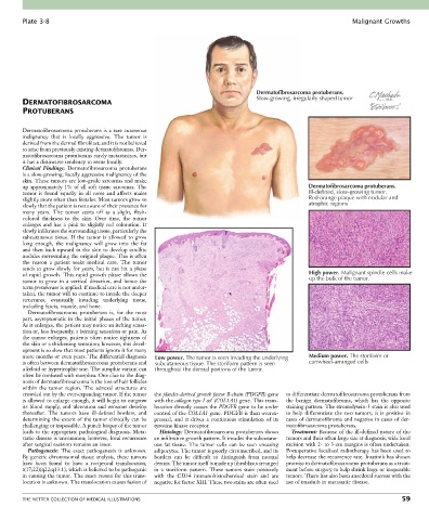
Plate 3-8                                                                                                Malignant Growths


                                                                                   Dermatofibrosarcoma protuberans.
        DERMATOFIBROSARCOMA                                                        Slow-growing, irregularly shaped tumor
        PROTUBERANS


        Dermatofibrosarcoma protuberans is a rare cutaneous
        malignancy  that  is  locally  aggressive.  The  tumor  is
        derived from the dermal fibroblast, and it is not believed
        to arise from previously existing dermatofibromas. Der-
        matofibrosarcoma protuberans rarely metastasizes, but
        it has a distinctive tendency to recur locally.
        Clinical  Findings:  Dermatofibrosarcoma  protuberans
        is a slow-growing, locally aggressive malignancy of the
        skin. These tumors are low-grade sarcomas and make
        up approximately 1% of all soft tissue sarcomas. The                                       Dermatofibrosarcoma protuberans.
        tumor  is  found  equally  in  all  races  and  affects  males                             Ill-defined, slow-growing tumor.
        slightly more often than females. Most tumors grow so                                      Red-orange plaque with nodular and
        slowly that the patient is not aware of their presence for                                 atrophic regions
        many  years.  The  tumor  starts  off  as  a  slight,  flesh-
        colored  thickness  to  the  skin.  Over  time,  the  tumor
        enlarges  and  has  a  pink  to  slightly  red  coloration.  It
        slowly infiltrates the surrounding tissue, particularly the
        subcutaneous  tissue.  If  the  tumor  is  allowed  to  grow
        long  enough,  the  malignancy  will  grow  into  the  fat
        and then back upward in the skin to develop satellite
        nodules surrounding the original plaque. This is often
        the  reason  a  patient  seeks  medical  care.  The  tumor
        tends to grow slowly for years, but it can hit a phase
        of  rapid  growth.  This  rapid  growth  phase  allows  the                                High power. Malignant spindle cells make
        tumor  to  grow  in  a  vertical  direction,  and  hence  the                              up the bulk of the tumor.
        term protuberans is applied. If medical care is not under-
        taken, the tumor will to continue to invade the deeper
        structures,  eventually  invading  underlying  tissue,
        including fascia, muscle, and bone.
          Dermatofibrosarcoma  protuberans  is,  for  the  most
        part, asymptomatic in the initial phases of the tumor.
        As it enlarges, the patient may notice an itching sensa-
        tion or, less frequently, a burning sensation or pain. As
        the tumor enlarges, patients often notice tightness of
        the skin or a thickening sensation; however, this devel-
        opment is so slow that most patients ignore it for many
        more months or even years. The differential diagnosis   Low power. The tumor is seen invading the underlying  Medium power. The storiform or
        is often between dermatofibrosarcoma protuberans and   subcutaneous tissue. The storiform pattern is seen  cartwheel-arranged cells
        a keloid or hypertrophic scar. The atrophic variant can   throughout the dermal portions of the tumor.
        often be confused with morphea. One clue to the diag-
        nosis of dermatofibrosarcoma is the loss of hair follicles
        within  the  tumor  region.  The  adnexal  structures  are
        crowded out by the ever-expanding tumor. If the tumor   the platelet-derived growth factor B-chain (PDGFB) gene   to differentiate dermatofibrosarcoma protuberans from
        is allowed to enlarge enough, it will begin to outgrow   with the collagen type I α1 (COL1A1) gene. This trans-  the  benign  dermatofibroma,  which  has  the  opposite
        its blood supply, and ulceration and erosions develop   location directly causes the PDGFB gene to be under   staining pattern. The stromolysein-3 stain is also used
        thereafter.  The  tumors  have  ill-defined  borders,  and   control of the COL1A1 gene. PDGFB is then overex-  to  help  differentiate  the  two  tumors;  it  is  positive  in
        determining the extent of the tumor clinically can be   pressed,  and  it  drives  a  continuous  stimulation  of  its   cases of dermatofibroma and negative in cases of der-
        challenging or impossible. A punch biopsy of the tumor   tyrosine kinase receptor.  matofibrosarcoma protuberans.
        leads to the appropriate pathological diagnosis. Meta-  Histology: Dermatofibrosarcoma protuberans shows   Treatment: Because of the ill-defined nature of the
        static disease is uncommon; however, local recurrence   an infiltrative growth pattern. It invades the subcutane-  tumors and their often large size at diagnosis, wide local
        after surgical excision remains an issue.  ous  fat  tissue.  The  tumor  cells  can  be  seen  encasing   excision with 2- to 3-cm margins is often undertaken.
          Pathogenesis:  The  exact  pathogenesis  is  unknown.   adipocytes. The tumor is poorly circumscribed, and its   Postoperative localized radiotherapy has been used to
        By genetic chromosomal tissue analysis, these tumors   borders  can  be  difficult  to  distinguish  from  normal   help decrease the recurrence rate. Imatinib has shown
        have  been  found  to  have  a  reciprocal  translocation,   dermis. The tumor itself is made up fibroblasts arranged   promise in dermatofibrosarcoma protuberans as a treat-
        t(17;22)(q22;q13.1), which is believed to be pathogenic   in  a  storiform  pattern.  These  tumors  stain  positively   ment before surgery to help shrink large or inoperable
        in causing the tumor. The exact reason for this trans-  with  the  CD34  immunohistochemical  stain  and  are   tumors. There has also been anecdotal success with the
        location is unknown. The translocation causes fusion of   negative for factor XIII. These two stains are often used   use of imatinib in metastatic disease.


        THE NETTER COLLECTION OF MEDICAL ILLUSTRATIONS                                                                           59