
Page 249 - Cardiac Nursing
P. 249
6/2
009
6/2
0/0
0/0
0:4
0:4
1
009
1
3
44.
q
44.
1-2
1-2
xd
3
xd
q
q
6 A
p
p
A
25
A
ara
ara
t
p
t
25
Pa
Pa
M
6 A
M
e 2
e 2
g
g
g
LWBK340-c10_
p
LWB
10_
LWB K34 0-c 10_ pp211-244.qxd 30/06/2009 10:46 AM Page 225 Aptara
p
K34
0-c
21
21
C HAPTER 1 0 / History Taking and Physical Examination 225
300
290
280
270 Lying Sitting Standing
260
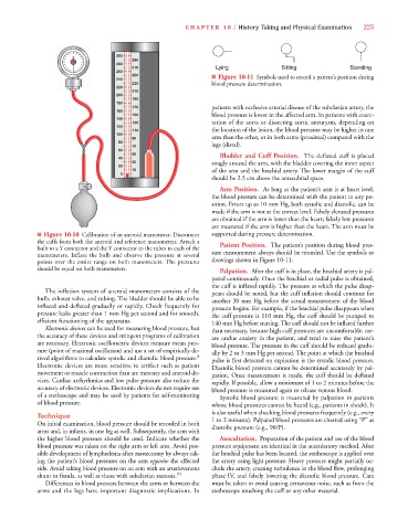
250 ■ Figure 10-11 Symbols used to record a patient’s position during
240
230 blood pressure determination.
220
210
200
190
180
170 patients with occlusive arterial disease of the subclavian artery, the
160
150 blood pressure is lower in the affected arm. In patients with coarc-
140
130 tation of the aorta or dissecting aortic aneurysm, depending on
120
110 the location of the lesion, the blood pressure may be higher in one
100
90 arm than the other, or in both arms (proximal) compared with the
80 legs (distal).
70
60
50
40 Bladder and Cuff Position. The deflated cuff is placed
30 snugly around the arm, with the bladder covering the inner aspect
20
10 of the arm and the brachial artery. The lower margin of the cuff
0
should be 2.5 cm above the antecubital space.
Arm Position. As long as the patient’s arm is at heart level,
the blood pressure can be determined with the patient in any po-
sition. Errors up to 10 mm Hg, both systolic and diastolic, can be
made if the arm is not at the correct level. Falsely elevated pressures
are obtained if the arm is lower than the heart; falsely low pressures
are measured if the arm is higher than the heart. The arm must be
■ Figure 10-10 Calibration of an aneroid manometer. Disconnect supported during pressure determination.
the cuffs from both the aneroid and reference manometers. Attach a Patient Position. The patient’s position during blood pres-
bulb to a Y connector and the Y connector to the tubes to each of the
manometers. Inflate the bulb and observe the pressure at several sure measurement always should be recorded. Use the symbols or
points over the entire range on both manometers. The pressures drawings shown in Figure 10-11.
should be equal on both manometers. Palpation. After the cuff is in place, the brachial artery is pal-
pated continuously. Once the brachial or radial pulse is obtained,
the cuff is inflated rapidly. The pressure at which the pulse disap-
The inflation system of aneroid manometers consists of the pears should be noted, but the cuff inflation should continue for
bulb, exhaust valve, and tubing. The bladder should be able to be another 30 mm Hg before the actual measurement of the blood
inflated and deflated gradually or rapidly. Check frequently for pressure begins. For example, if the brachial pulse disappears when
pressure leaks greater than 1 mm Hg per second and for smooth, the cuff pressure is 110 mm Hg, the cuff should be pumped to
efficient functioning of the apparatus. 140 mm Hg before starting. The cuff should not be inflated further
Electronic devices can be used for measuring blood pressure, but than necessary, because high cuff pressures are uncomfortable, cre-
the accuracy of these devices and stringent programs of calibration ate undue anxiety in the patient, and tend to raise the patient’s
are necessary. Electronic oscillometric devices measure mean pres- blood pressure. The pressure in the cuff should be reduced gradu-
sure (point of maximal oscillation) and use a set of empirically de- ally by 2 to 3 mm Hg per second. The point at which the brachial
rived algorithms to calculate systolic and diastolic blood pressure. 6 pulse is first detected on expiration is the systolic blood pressure.
Electronic devices are more sensitive to artifact such as patient Diastolic blood pressure cannot be determined accurately by pal-
movement or muscle contraction than are mercury and aneroid de- pation. Once measurement is made, the cuff should be deflated
vices. Cardiac arrhythmias and low pulse pressure also reduce the rapidly. If possible, allow a minimum of 1 to 2 minutes before the
accuracy of electronic devices. Electronic devices do not require use blood pressure is measured again to release venous blood.
of a stethoscope and may be used by patients for self-monitoring Systolic blood pressure is measured by palpation in patients
of blood pressure. whose blood pressures cannot be heard (e.g., patients in shock). It
is also useful when checking blood pressures frequently (e.g., every
Technique 1 to 2 minutes). Palpated blood pressures are charted using “P” as
On initial examination, blood pressure should be recorded in both diastolic pressure (e.g., 90/P).
arms and, in infants, in one leg as well. Subsequently, the arm with
the higher blood pressure should be used. Indicate whether the Auscultation. Preparation of the patient and use of the blood
blood pressure was taken on the right arm or left arm. Avoid pos- pressure equipment are identical in the auscultatory method. After
sible development of lymphedema after mastectomy by always tak- the brachial pulse has been located, the stethoscope is applied over
ing the patient’s blood pressures on the arm opposite the affected the artery using light pressure. Heavy pressure might partially oc-
side. Avoid taking blood pressure on an arm with an arteriovenous clude the artery, creating turbulence in the blood flow, prolonging
shunt or fistula, as well as those with subclavian stenosis. 24 phase IV, and falsely lowering the diastolic blood pressure. Care
Differences in blood pressure between the arms or between the must be taken to avoid causing extraneous noise, such as from the
arms and the legs have important diagnostic implications. In stethoscope touching the cuff or any other material.