
Page 257 - Cardiac Nursing
P. 257
LWBK340-c10_p211-244.qxd 30/06/2009 10:47 AM Page 233 Aptara
C HAP TE R 1 0 / History Taking and Physical Examination 233
The intensity of the S 1 depends on leaflet mobility, position of
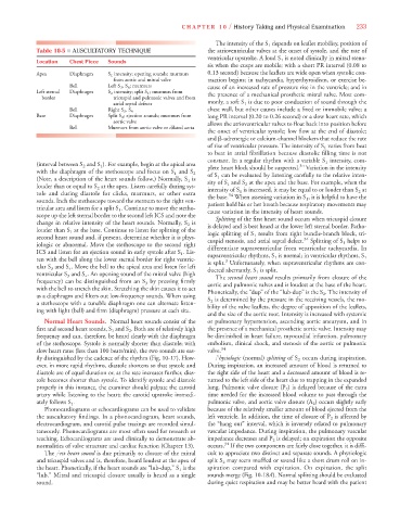
Table 10-5 ■ AUSCULTATORY TECHNIQUE the atrioventricular valves at the onset of systole, and the rate of
ventricular upstroke. A loud S 1 is noted clinically in mitral steno-
Location Chest Piece Sounds
sis when the cusps are mobile; with a short PR interval (0.08 to
Apex Diaphragm S 1 intensity; opening sounds; murmurs 0.13 second) because the leaflets are wide open when systolic con-
from aortic and mitral valve traction begins; in tachycardia, hyperthyroidism, or exercise be-
Bell Left S 3 , S 4 ; murmurs cause of an increased rate of pressure rise in the ventricle; and in
Left sternal Diaphragm S 2 intensity; split S 1 ; murmurs from
border tricuspid and pulmonic valves and from the presence of a mechanical prosthetic mitral valve. Most com-
atrial septal defects monly, a soft S 1 is due to poor conduction of sound through the
Bell Right S 3 , S 4 chest wall, but other causes include a fixed or immobile valve; a
Base Diaphragm Split S 2 ; ejection sounds; murmurs from long PR interval (0.20 to 0.26 second) or a slow heart rate, which
aortic valve allows the atrioventricular valves to float back into position before
Bell Murmurs from aortic valve or dilated aorta
the onset of ventricular systole; low flow at the end of diastole;
and -adrenergic or calcium-channel blockers that reduce the rate
of rise of ventricular pressure. The intensity of S 1 varies from beat
to beat in atrial fibrillation because diastolic filling time is not
constant. In a regular rhythm with a variable S 1 intensity, com-
(interval between S 2 and S 1 ). For example, begin at the apical area 34
plete heart block should be suspected. Variation in the intensity
with the diaphragm of the stethoscope and focus on S 1 and S 2 of S 1 can be evaluated by listening carefully to the relative inten-
(Note, a description of the heart sounds follow.) Normally, S 1 is sity of S 1 and S 2 at the apex and the base. For example, when the
louder than or equal to S 2 at the apex. Listen carefully during sys- intensity of S 1 is increased, it may be equal to or louder than S 2 at
tole and during diastole for clicks, murmurs, or other extra the base. When assessing variation in S 1 , it is helpful to have the
34
sounds. Inch the stethoscope toward the sternum to the right ven- patient hold his or her breath because respiratory movements may
tricular area and listen for a split S 1 . Continue to move the stetho- cause variation in the intensity of heart sounds.
scope up the left sternal border to the second left ICS and note the Splitting of the first heart sound occurs when tricuspid closure
change in relative intensity of the heart sounds. Normally, S 2 is is delayed and is best heard at the lower left sternal border. Patho-
louder than S 1 at the base. Continue to listen for splitting of the logic splitting of S 1 results from right bundle-branch block, tri-
second heart sound and, if present, determine whether it is phys- cuspid stenosis, and atrial septal defect. 34 Splitting of S 1 helps to
iologic or abnormal. Move the stethoscope to the second right differentiate supraventricular from ventricular tachycardia. In
ICS and listen for an ejection sound in early systole after S 1 . Lis-
supraventricular rhythms, S 1 is normal; in ventricular rhythms, S 1
ten with the bell along the lower sternal border for right ventric- is split. Unfortunately, when supraventricular rhythms are con-
3
ular S 3 and S 4 . Move the bell to the apical area and listen for left ducted aberrantly, S 1 is split.
ventricular S 3 and S 4 . An opening sound of the mitral valve (high The second heart sound results primarily from closure of the
frequency) can be distinguished from an S 3 by pressing firmly aortic and pulmonic valves and is loudest at the base of the heart.
with the bell to stretch the skin. Stretching the skin causes it to act Phonetically, the “dup” of the “lub-dup” is the S 2 . The intensity of
as a diaphragm and filters out low-frequency sounds. When using S 2 is determined by the pressure in the receiving vessels, the mo-
a stethescope with a tunable diaphragm one can alternate listen- bility of the valve leaflets, the degree of apposition of the leaflets,
ing with light (bell) and firm (diaphragm) pressure at each site.
and the size of the aortic root. Intensity is increased with systemic
Normal Heart Sounds. Normal heart sounds consist of the or pulmonary hypertension, ascending aortic aneurysm, and in
first and second heart sounds, S 1 and S 2 . Both are of relatively high the presence of a mechanical prosthetic aortic valve. Intensity may
frequency and can, therefore, be heard clearly with the diaphragm be diminished in heart failure, myocardial infarction, pulmonary
of the stethoscope. Systole is normally shorter than diastole; with embolism, clinical shock, and stenosis of the aortic or pulmonic
slow heart rates (less than 100 beats/min), the two sounds are eas- valve. 34
ily distinguished by the cadence of the rhythm (Fig. 10-17). How- Physiologic (normal) splitting of S 2 occurs during inspiration.
ever, in more rapid rhythms, diastole shortens so that systole and During inspiration, an increased amount of blood is returned to
diastole are of equal duration or, as the rate increases further, dias- the right side of the heart and a decreased amount of blood is re-
tole becomes shorter than systole. To identify systole and diastole turned to the left side of the heart due to trapping in the expanded
properly in this instance, the examiner should palpate the carotid lung. Pulmonic valve closure (P 2 ) is delayed because of the extra
artery while listening to the heart; the carotid upstroke immedi- time needed for the increased blood volume to pass through the
A
ately follows S 1 . pulmonic valve, and aortic valve closure (A 2 ) occurs slightly early
Phonocardiograms or echocardiograms can be used to validate because of the relatively smaller amount of blood ejected from the
the auscultatory findings. In a phonocardiogram, heart sounds, left ventricle. In addition, the time of closure of P 2 is affected by
electrocardiogram, and carotid pulse tracings are recorded simul- the “hang out” interval, which is inversely related to pulmonary
taneously. Phonocardiograms are most often used for research or vascular impedance. During inspiration, the pulmonary vascular
teaching. Echocardiograms are used clinically to demonstrate ab- impedance decreases and P 2 is delayed; on expiration the opposite
34
normalities of valve structure and cardiac function (Chapter 13). occurs. If the two components are fairly close together, it is diffi-
The first heart sound is due primarily to closure of the mitral cult to appreciate two distinct and separate sounds. A physiologic
and tricuspid valves and is, therefore, heard loudest at the apex of split S 2 may seem muffled or sound like a short drum roll on in-
the heart. Phonetically, if the heart sounds are “lub-dup,” S 1 is the spiration compared with expiration. On expiration, the split
8
8
“lub.” Mitral and tricuspid closure usually is heard as a single sounds merge (Fig. 10-18A). Normal splitting should be evaluated
sound. during quiet respiration and may be better heard with the patient