
Page 254 - Cardiac Nursing
P. 254
0/0
6/2
0/0
3
3
6/2
1
0:4
1
009
009
xd
1-2
1-2
21
p
21
44.
q
xd
q
44.
q
A
p
A
30
30
p
ara
ara
t
p
t
e 2
M
M
7 A
0:4
7 A
Pa
g
e 2
g
Pa
g
LWB
K34
LWB K34 0-c 10_ pp211-244.qxd 30/06/2009 10:47 AM Page 230 Aptara
LWBK340-c10_
10_
0-c
p
230 P A R T III / Assessment of Heart Disease
6
a b c edema, or increased capillary permeability. In the cardiac patient,
b peripheral edema frequently occurs because of sodium and water
c a
retention and right-sided heart failure. Bilateral edema of the lower
extremities suggests a systemic etiology; unilateral edema is usually
the result of a local etiology. A weight gain of 10 lb (indicative of
A abc < 180 ° abc > 195 ° 5 L of extracellular fluid volume) precedes visible edema in most
patients. Interstitial edema occurs in the most dependent part of
the body, its location varying with the patient’s posture. With sit-
ting or standing, edema develops in the lower extremities. With
IPD bedrest, edema forms in the sacrum. Because the distribution of
DPD DPD IPD
edema fluid varies with position, daily weights provide the best se-
rial assessment of edema. Pitting edema is a depression in the skin
from pressure. To demonstrate the presence of pitting edema, the
nurse presses firmly with his or her thumb over a bony surface such
B DPD < IPD DPD > IPD as the sacrum, medial malleolus, the dorsum of each foot, and the
shins. When the thumb is withdrawn, an indentation persists for a
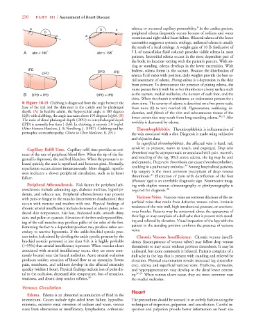
■ Figure 10-15 Clubbing is diagnosed from the angle between the short time. The severity of edema is described on a five-point scale,
base of the nail and the skin next to the cuticle and by phalangeal from none (0) to very marked (4). Pigmentation, reddening, g in-
g
depth. (A) In healthy adults, the hyponychial angle is 180 degrees duration, and fibrosis of the skin and subcutaneous tissues of the
t
t
t
t
(left); with clubbing, the angle increases above 195 degrees (right). (B) lower extremities may result from long-standing edema. 30,31 Skin
The ratio of distal phalangeal depth (DPD) to interphalangeal depth mobility is decreased by edema.
t
(IPD) is normally less then 1 (left). In clubbing, it exceeds 1.0 (t t right).
t
(After Hansen-Flaschen, J. & Nordberg, J. [1987]. Clubbing and hy- Thrombophlebitis. Thrombophlebitis is inflammation of
pertrophic osteoarthropathy. Clinics in Chest Medicine, 8, 291.) the vein associated with a clot. Diagnosis is made using subjective
and objective data.
In superficial thrombophlebitis, the affected vein is hard, red,
sensitive to pressure, warm to touch, and engorged. Deep vein
Capillary Refill Time. Capillary refill time provides an esti-
mate of the rate of peripheral blood flow. When the tip of the fin- thrombosis may be asymptomatic or associated with pain, warmth,
gernail is depressed, the nail bed blanches. When the pressure is re- and mottling of the leg. With severe edema, the leg may be cool
leased quickly, the area is reperfused and becomes pink. Normally, and cyanotic. Deep vein thrombosis can cause thromboembolism,
32
reperfusion occurs almost instantaneously. More sluggish reperfu- resulting in a pulmonary embolus. Among hospitalized patients,
sion indicates a slower peripheral circulation, such as in heart hip surgery is the most common precipitant of deep venous
31
failure. thrombosis. Elicitation of pain with dorsiflexion of the foot
(Homans’ sign) is an unreliable diagnostic sign. Noninvasive imag-
Peripheral Atherosclerosis. Risk factors for peripheral ath- ing with duplex venous ultrasonography or plethysmography is
erosclerosis include advancing age, diabetes mellitus, hyperlipi- required for diagnosis. 32
demia, and tobacco use. Peripheral atherosclerosis may present
with pain or fatigue in the muscles (intermittent claudication) that Varicose Veins. Varicose veins are tortuous dilations of the su-
occurs with exercise and resolves with rest. Physical findings of perficial veins that result from defective venous valves, intrinsic
chronic arterial insufficiency include decreased or absent pulses, re- weakness of the vein wall, high intraluminal pressure, or arteriove-
duced skin temperature, hair loss, thickened nails, smooth shiny nous fistulas. Patients may be concerned about the appearance of
skin, and pallor or cyanosis. Elevation of the feet and repeated flex- their legs or may complain of a dull ache that is present with stand-
ing of the calf muscles may produce pallor of the soles of the feet. ing and relieved by elevation. Visual inspection of the legs with the
Returning the feet to a dependent position may produce rubor sec- patient in the standing position confirms the presence of varicose
ondary to reactive hyperemia. If the ankle–brachial systolic pres- veins.
sure index (calculated by dividing the ankle systolic pressure by the Chronic Venous Insufficiency. Chronic venous insuffi-
brachial systolic pressure) is less than 0.8, it is highly probable ciency (incompetence of venous valves) may follow deep venous
( 95%) that arterial insufficiency is present. When vascular ulcers thrombosis or may occur without previous thrombosis. It may be
associated with arterial insufficiency occur, they are more com- unilateral, but more commonly is bilateral. Patients complain of a
monly located near the lateral malleolus. Acute arterial occlusion dull ache in the legs that is present with standing and relieved by
produces sudden cessation of blood flow to an extremity. Severe elevation. Physical examination reveals increased leg circumfer-
pain, numbness, and coldness develop in the affected extremity ence, edema, and superficial varicose veins. Erythema, dermatitis,
quickly (within 1 hour). Physical findings include loss of pulse dis- and hyperpigmentation may develop in the distal lower extrem-
tal to the occlusion, decreased skin temperature, loss of sensation, ity. 30,31 When venous ulcers occur, they are more common near
weakness, and absent deep tendon reflexes. 30 the medial malleolus.
Venous Circulation
Heart
Edema. Edema is an abnormal accumulation of fluid in the
interstitium. Causes include right-sided heart failure, hypoalbu- The precordium should be assessed in an orderly fashion using the
minemia, excessive renal retention of sodium and water, venous techniques of inspection, palpation, and auscultation. Careful in-
stasis from obstruction or insufficiency, lymphedema, orthostatic spection and palpation provide better information on heart size