
Page 258 - Cardiac Nursing
P. 258
6/2
6/2
009
3
0/0
0/0
009
0:4
7 A
7 A
1
1
0:4
3
p
1-2
1-2
21
21
p
44.
q
xd
xd
44.
q
q
t
t
ara
p
p
p
ara
LWB
LWBK340-c10_
LWB K34 0-c 10_ pp211-244.qxd 30/06/2009 10:47 AM Page 234 Aptara
10_
0-c
K34
g
g
g
Pa
M
M
Pa
34
A
A
34
e 2
e 2
234 P A R T III / Assessment of Heart Disease
insp insp
S 1 S 1 S 1 S 1 S 3 S 1 S 3 S 1 S 3
2
2
A A P 2 S 2 A P 2 S 2 S 2 S 2
■ Figure 10-19 An S 3 gallop immediately follows the S 2 .
exp insp exp
“lub-dup-ta” cadence (Fig. 10-19). Using the bell of the stetho-
scope, listen for a left ventricular S 3 over the apex of the heart; for
S 1 S 1 S 1
2
2
B P A 2 S 2 P A 2 a right ventricular S 3 , listen over the lower left sternal border. By
having the patient in the left lateral position, the apex is brought
■ Figure 10-18 Splitting of the S 2 . (A) Physiologic splitting. Dur-
forward against the chest wall, making the left ventricular S 3
ing inspiration (insp), the P 2 sound is delayed. (B) Paradoxical split-
A
ting. During expiration (exp), A 2 is delayed. louder and, therefore, easier to hear.
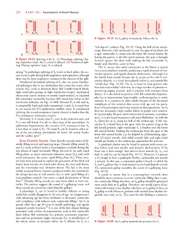
The S 4 occurs after atrial contraction as the blood is ejected
3
sitting. In pathologic splitting of S 2 (wide or fixed splits), the sec- into a noncompliant ventricle, producing a rapid elevation of ven-
tricular pressure, and signals diastolic dysfunction. Although it is
ond sound is split during both inspiration and expiration, although
the fourth heart sound, because the S 4 occurs at the end of ven-
there may be some respiratory variation in the amount of the split.
tricular diastole, it is heard immediately before S 1 and sounds like
Paradoxical (abnormal) splitting of S 2 also can occur. Paradox-
“ta-lub-dup” (Fig. 10-20). The S 4 is heard in most patients who
ical splitting is due to any mechanism that causes late aortic valve
have had a myocardial infarction, in a large number of patients ex-
closure (A 2 ), such as electrical delay (left bundle-branch block,
periencing angina pectoris, and in patients with coronary heart
right ventricular pacing, or right ventricular ectopy), mechanical
disease. It is also heard in patients with left ventricular hypertro-
obstruction (aortic stenosis or systolic hypertension), or impaired
phy due to hypertension, hypertrophic cardiomyopathy, or aortic
left ventricular contractile function (left ventricular failure or left
stenosis. It is common in older adults because of the decreased
B A
ventricular ischemia; see Fig. 10-18B). Because P 2 is soft and A 2
compliance of the ventricle that occurs with age and the preva-
is comparably loud and easily transmitted, a split S 2 is heard best
in the second left ICS (pulmonary outflow tract). In paradoxical lence of hypertension and aortic stenosis in this population. An S 4
does not necessarily imply cardiac failure in people with ventricu-
splitting, the second component (aortic closure) is louder than the
lar hypertrophy. Because atrial contraction is necessary to produce
first component (pulmonic closure).
an S 4 , it is not heard in patients with atrial fibrillation. As with the
Normally, A 2 is louder than P 2 , even in the pulmonic area, and
S 3 , listen for an S 4 using the bell of the stethoscope. A left ven-
P 2 is not well heard, if at all, in other areas of the precordium. In
tricular S 4 is heard best at the apex, with the patient lying in the
pulmonary hypertension, the intensity of P 2 increases so that A 2
left lateral position; right ventricular S 4 is loudest over the lower
is less than or equal to P 2 . The loud P 2 can be heard in other ar-
left sternal border. Inching the stethoscope from the apex to the
eas of the precordium, particularly the lower left sternal border
and the cardiac apex. 8 lower left sternal border can be helpful in differentiating right-
and left-sided sounds. Left-sided sounds fade and right-sided
Extra Diastolic Sounds. Extra diastolic sounds consist of di- sounds get louder as the stethoscope approaches the sternum.
A quadruple rhythm may be heard in patients with severe car-
astolic filling sounds and opening snaps. Diastolic filling sounds (S 3
and S 4 ) occur as blood enters a noncompliant ventricle during the diac failure and both systolic and diastolic dysfunctions. If the
two phases of rapid ventricular filling: the end of the early rapid heart rate is slow enough, four distinct heart sounds (S 1 , S 2 , and
filling phase, as active ventricular relaxation ceases (S 3 ); and, with both S 3 and S 4 ) can be heard (Fig. 10-21). However, if a patient
atrial contraction, the active, rapid filling phase (S 4 ). Three theo- is ill enough to have a quadruple rhythm, tachycardia also usually
ries have been proposed to explain the generation of the third and is present. In this case, a summation gallop is heard, in which the
fourth heart sounds: the mitral valve theory, the chest wall theory, S 3 and S 4 gallops fuse in mid-diastole to one loud diastolic sound.
and the ventricular wall vibration theory. The last is the most The summation gallop resembles the sound of a galloping horse
widely accepted theory. Sound is produced within the ventricle by (Fig. 10-22).
the abrupt decrease in wall motion (S 3 ) or with rapid filling of a It stands to reason that in a noncompliant ventricle there
noncompliant ventricle that causes a rapid deceleration of blood should be more resistance to active ventricular filling than to pas-
flow. 35 Diastolic filling sounds can arise from either or both ven- sive ventricular filling; therefore, an S 4 gallop should be generated
tricles. The cadence suggests the sound of a galloping horse, and more easily than an S 3 gallop. Therefore, one would expect all pa-
these sounds are sometimes called diastolic gallops. tients with normal sinus rhythm who have an S 3 gallop to have an
A physiologic S 3 S can be heard in healthy children or young S 4 gallop as well. However, patients with normal sinus rhythm fre-
adults but usually disappears by 40 years of age. Its disappearance quently have only an S 3 . The cause for this finding is unknown,
with advancing age has been attributed to decreased ventricular
3
wall compliance with reduced early ventricular filling. An S 3 in
people older than age 40 years is usually pathologic and signals
impaired systolic function. 35 It is one of the first clinical findings
associated with cardiac decompensation, such as left ventricular
heart failure (left ventricular S 3 ), primary pulmonary hyperten- S S 1 S S S 1 S S S 1 S
4
4
4
sion and cor pulmonale (right ventricular S 3 ), or insufficiency of 2 2 2
the mitral, aortic, or tricuspid valves. An S 3 follows the S 2 in a ■ Figure 10-20 An S 4 gallop immediately precedes the S 1 .