
Page 252 - Cardiac Nursing
P. 252
21
21
p
g
p
g
44.
q
44.
1-2
1-2
e 2
p
p
t
ara
t
p
28
e 2
28
A
A
0:4
7 A
0:4
1
1
7 A
Pa
g
Pa
M
M
009
xd
3
xd
q
q
3
6/2
009
6/2
0/0
0/0
K34
0-c
10_
LWBK340-c10_
ara
LWB K34 0-c 10_ pp211-244.qxd 30/06/2009 10:47 AM Page 228 Aptara
LWB
228 P A R T III / Assessment of Heart Disease
blood pressure, increased pulse, or symptoms in the sitting
position presage similar events in the erect position. Often, the
change in blood pressure does not meet the criteria for orthosta-
sis, but it is accompanied by a significant change in heart rate or
associated symptoms, or both. These circumstances identify peo-
ple at risk and should prompt further investigation by the cardiac
nurse of the patient’s present volume status and vasodilatory or
cardioinhibitory drug regimen.
The presence of intravascular volume depletion (such as with
diuretic therapy) should be suspected when, in response to sit-
ting or standing, the heart rate increases and the systolic pressure
decreases by 15 mm Hg and the diastolic blood pressure drops
by 10 mm Hg. 26 It is difficult to differentiate intravascular vol-
ume loss from inadequate vasoconstrictor mechanisms solely by
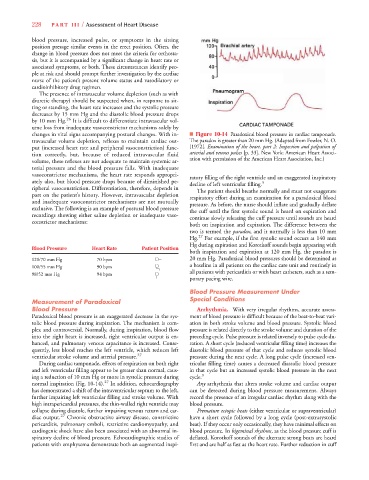
changes in vital signs accompanying postural changes. With in- ■ Figure 10-14 Paradoxical blood pressure in cardiac tamponade.
travascular volume depletion, reflexes to maintain cardiac out- The paradox is greater than 20 mm Hg. (Adapted from Fowler, N. O.
put (increased heart rate and peripheral vasoconstriction) func- [1972]. Examination of the heart, part 2: Inspection and palpation of
tion correctly, but, because of reduced intravascular fluid arterial and venous pulses [p. 33]. New York: American Heart Associ-
volume, these reflexes are not adequate to maintain systemic ar- ation with permission of the American Heart Association, Inc.)
terial pressure and the blood pressure falls. With inadequate
vasoconstrictor mechanisms, the heart rate responds appropri- ratory filling of the right ventricle and an exaggerated inspiratory
ately also, but blood pressure drops because of diminished pe- decline of left ventricular filling. 5
ripheral vasoconstriction. Differentiation, therefore, depends in The patient should breathe normally and must not exaggerate
part on the patient’s history. However, intravascular depletion respiratory effort during an examination for a paradoxical blood
and inadequate vasoconstrictor mechanisms are not mutually pressure. As before, the nurse should inflate and gradually deflate
exclusive. The following is an example of postural blood pressure the cuff until the first systolic sound is heard on expiration and
recordings showing either saline depletion or inadequate vaso- continue slowly releasing the cuff pressure until sounds are heard
constrictor mechanisms:
both on inspiration and expiration. The difference between the
two is termed the paradox, and it normally is less than 10 mm
Hg. 27 For example, if the first systolic sound occurs at 140 mm
Hg during expiration and Korotkoff sounds begin appearing with
Blood Pressure Heart Rate Patient Position
both inspiration and expiration at 120 mm Hg, the paradox is
120/70 mm Hg 70 bpm 20 mm Hg. Paradoxical blood pressures should be determined as
100/55 mm Hg 90 bpm a baseline in all patients on the cardiac care unit and routinely in
all patients with pericarditis or with heart catheters, such as a tem-
98/52 mm Hg 94 bpm
porary pacing wire.
Blood Pressure Measurement Under
Special Conditions
Measurement of Paradoxical
Blood Pressure Arrhythmia. With very irregular rhythms, accurate assess-
Paradoxical blood pressure is an exaggerated decrease in the sys- ment of blood pressure is difficult because of the beat-to-beat vari-
tolic blood pressure during inspiration. The mechanism is com- ation in both stroke volume and blood pressure. Systolic blood
plex and controversial. Normally, during inspiration, blood flow pressure is related directly to the stroke volume and duration of the
into the right heart is increased, right ventricular output is en- preceding cycle. Pulse pressure is related inversely to pulse cycle du-
hanced, and pulmonary venous capacitance is increased. Conse- ration. A short cycle (reduced ventricular filling time) increases the
quently, less blood reaches the left ventricle, which reduces left diastolic blood pressure of that cycle and reduces systolic blood
ventricular stroke volume and arterial pressure. 27 pressure during the next cycle. A long pulse cycle (increased ven-
During cardiac tamponade, effects of respiration on both right tricular filling time) causes a decreased diastolic blood pressure
and left ventricular filling appear to be greater than normal, caus- in that cycle but an increased systolic blood pressure in the next
ing a reduction of 10 mm Hg or more in systolic pressure during cycle. 3
normal inspiration (Fig. 10-14). 27 In addition, echocardiography Any arrhythmia that alters stroke volume and cardiac output
has demonstrated a shift of the intraventricular septum to the left, can be detected during blood pressure measurement. Always
further impairing left ventricular filling and stroke volume. With record the presence of an irregular cardiac rhythm along with the
high intrapericardial pressures, the thin-walled right ventricle may blood pressure.
collapse during diastole, further impairing venous return and car- Premature ectopic beats (either ventricular or supraventricular)
diac output. 27 Chronic obstructive airway disease, constrictive have a short cycle followed by a long cycle (post-extrasystolic
pericarditis, pulmonary emboli, restrictive cardiomyopathy, and beat). If they occur only occasionally, they have minimal effects on
cardiogenic shock have also been associated with an abnormal in- blood pressure. In bigeminal rhythms, as the blood pressure cuff is
spiratory decline of blood pressure. Echocardiographic studies of deflated, Korotkoff sounds of the alternate strong beats are heard
patients with emphysema demonstrate both an augmented inspi- first and are half as fast as the heart rate. Further reduction in cuff