
Page 256 - Cardiac Nursing
P. 256
p
p
p
t
t
21
1-2
1-2
p
21
e 2
ara
g
32
g
Pa
Pa
e 2
32
ara
A
p
A
44.
1
1
009
6/2
009
0:4
M
M
7 A
0:4
7 A
6/2
q
xd
q
44.
q
xd
0/0
0/0
3
g
3
0-c
0-c
10_
10_
K34
LWB
LWBK340-c10_ pp211-244.qxd 30/06/2009 10:47 AM Page 232 Aptara
K34
LWB
232 P A R T III / Assessment of Heart Disease
Palpation The parts of the stethoscope are the ear pieces, tubing, and
Movement that was not visible on inspection may be detected by chest pieces. The ear pieces should fit comfortably into the ear
palpation. All areas should be palpated using either the ball of the canal and be snug enough so that extraneous sound cannot enter.
palm (at the base of the fingers) or the fingertips. In general, the They also must be kept free of ear wax. Double tubing with a
palm surface is more sensitive to thrills (vibrations), whereas fin- small internal diameter (3 mm) should extend from the ear pieces
gertips are more sensitive to pulsations. Thrills indicate turbulence to the chest pieces. In addition, the tubing should be reasonably
of blood flow and are associated with murmurs. Impulses are de- short (25 to 30 cm) so the sound is not diluted and should be
scribed in terms of location, size, amplitude, duration, and time in thick to minimize room noise. 33
the cardiac cycle (systole or diastole). To facilitate measurement of There are two classic types of chest pieces, the diaphragm
the horizontal location in centimeters from the MCL, or the size and the bell. The diaphragm, which brings out higher frequen-
of the impulse, it is helpful for the examiner to measure his or her cies and filters out the lower ones, is useful for listening to the
hand and use it as a “ruler.” For example, the distance from the tip first and second heart sounds (S 1 and S 2 ), high-frequency mur-
of the finger to the first joint, the second joint, and the third joint murs, and lung sounds. The diaphragm should be pressed
can be used. firmly against the chest wall. The bell filters out high-frequency
Assess the apex impulse for location, size, amplitude, and du- sounds and accentuates the low-frequency ones. Diastolic fill-
ration. The apex impulse is, by definition, the furthest point left- ing sounds and the low-frequency murmurs of mitral and tri-
ward and downward at which a cardiac pulsation can be seen or cuspid stenosis are heard best with the bell. 33 The bell should
3
felt. The normal apex impulse is felt as a light tap, extending over rest lightly on the chest; if firm pressure is applied, the skin be-
3 cm or less. The apex impulse is felt immediately after the first comes taut and acts like a diaphragm. When auscultating heart
heart sound and lasts halfway through systole. An impulse that is sounds, the nurse stands on the patient’s right side so that, as he
diffuse (felt over two ICSs), increased in amplitude, or laterally or or she places the bell of the stethoscope on the patient’s chest,
inferiorly displaced suggests increased volume load and left ven- the chest piece is balanced. Because the bell does not have to be
tricular dilatation, such as occurs in mitral insufficiency or left held in place, the possibilities of creating extraneous sounds and
ventricular failure. An impulse that is sustained, enlarged, and, filtering out low frequencies are reduced. Some stethoscopes
sometimes, laterally displaced suggests obstruction to outflow have a single chestpiece with tunable diaphragm. Very light skin
with increased ventricular pressure load and concentric hypertro- contact is used to listen to low-frequency sounds and firm pres-
phy of the muscle, such as occurs in aortic stenosis or systemic sure is used to listen to high-frequency sounds.
6
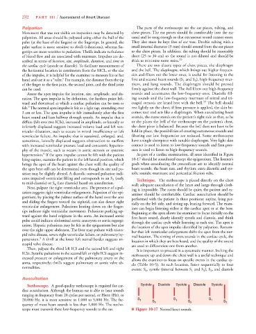
hypertension. If the apex impulse cannot be felt with the patient As part of a cardiac examination, all areas identified in Figure
lying supine, examine the patient in the left lateral position, which 10-17 should be auscultated except the epigastrium. The listener’s
brings the apex of the heart against the chest wall; the quality of goals when auscultating the precordium are to identify normal
the apex beat still can be determined even though its size and po- heart sounds, the heart rate, and rhythm; extra diastolic and sys-
sition may be slightly altered. A diastolic outward pulsation indi- tolic sounds; murmurs; and pericardial friction rubs.
cates impaired ventricular filling and corresponds to an S 3 (early Technique. The stethoscope is placed directly on the chest
to mid-diastole) or S 4 (late diastole) heard on auscultation. wall; adequate auscultation of the heart and lungs through cloth-
Next, palpate the right ventricular area. The presence of a pul- ing is impossible. The room should be quiet; the patient and ex-
sation suggests right ventricular enlargement. Palpation of the epi- aminer should be comfortable. Cardiac auscultation should be
gastrium, by placing the palmar surface of the hand over the area performed with the patient in three positions: supine, lying par-
and sliding the fingers toward the xiphoid, can also detect right tially on the left side, and sitting up, leaning forward. The exam-
ventricular enlargement. Pulsations beating down on the finger- iner can begin listening either at the cardiac apex or at the base.
tips indicate right ventricular movement. Pulsations pushing up- Beginning at the apex allows the examiner to focus initially on the
ward against the hand originate in the aorta. An increased aortic first heart sound, clearly identify systole and diastole, and think
pulse could indicate abdominal aortic aneurysm or aortic regurgi- through the cardiac cycle while listening at each site. The apex is
tation. Hepatic pulsations may be felt in the epigastrium but also the location of the apex impulse identified by palpation. Remem-
over the right upper abdomen. The liver may pulsate with tricus- ber that left ventricular enlargement shifts the apex from the nor-
pid valve disease, severe right ventricular failure, or pulmonary hy- mal location. The timing of extra sounds in the cardiac cycle, the
3
pertension. A thrill at the lower left sternal border suggests tri- location in which they are best heard, and the quality of the sound
cuspid valve disease. are used to differentiate one from another.
Then, palpate the third left ICS and the second left and right It is important to proceed in a systematic manner. Inching the
ICSs. Systolic pulsations in the second left or right ICS suggest in- stethoscope up and down the chest wall is a useful technique and
creased pressure or enlargement of the pulmonary artery or the allows the examiner to focus on specific events in the cardiac cy-
aorta, respectively; thrills suggest pulmonary or aortic valve ab- cle (Table 10-5). At each location, listen sequentially to four
normalities.
events: S 1 , systole (interval between S 1 and S 2 ), S 2 , and diastole
Auscultation
Stethoscope. A good-quality stethoscope is required for car- Systole Diastole Systole Systole
diac auscultation. Although the human ear is able to hear sounds
ranging in frequency from 20 cycles per second, or Hertz (Hz), to
20,000 Hz, it is most sensitive to 1,000 to 5,000 Hz. The fre- S 1 S 2 S 1 S 2 S 1 S 2
quency of most heart sounds is less than 1,000 Hz. The stetho-
scope must transmit these low-frequency sounds to the ear. ■ Figure 10-17 Normal heart sounds.