
Page 303 - Cardiac Nursing
P. 303
6/2
6/2
79
009
1
1
009
A
3
xd
xd
3
0/0
0/0
3
g
g
Pa
g
79
e 2
e 2
Pa
2 A
0:4
0:4
2 A
M
M
M
q
t
t
ara
p
p
p
p
ara
LWB
LWBK340-c13_
LWB K34 0-c 13_ pp277-290.qxd 30/06/2009 10:42 AM Page 279 Aptara
K34
ara
13_
0-c
A
7-2
7-2
90.
q
q
90.
27
p
27
C HAPTER 1 3 / Echocardiography 279
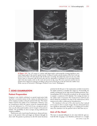
■ Figure 13-3 Top: 2-D image in a patient with hypertrophic cardiomyopathy showing significant asym-
metric hypertrophy of the anterior septum (arrow). Evidence of systolic anterior motion of the mitral valve ap-
t
paratus (arrow) with 2-D echo (bottom, left). In this same 2-D image, the dashed line represents the area of the
t
narrow beam of ultrasound with M-mode echo and that information is displayed in the next image (bottom,
t
t
x
right) with time (cardiac cycle) on the x-axis and motion on the y-axis. The arrow represents systolic anterior
x
motion of the mitral valve apparatus resulting in obstruction of blood flow in the left ventricular outflow tract.
(Echo courtesy of University of Washington Medical Center, Seattle, Washington.)
position for the first part of the examination and then is moved to
ECHO EXAMINATION the supine position to complete the image set. Occasionally, the
patient is positioned in the right lateral decubitus position. Elec-
Patient Preparation trocardiogram (ECG) electrodes are placed on the patient’s skin
to acquire a continuous ECG rhythm. The timing of events to
Imaging is most ideally performed on special examination beds the cardiac cycle is important. The blood pressure and heart rate
that have a removable section of the mattress to allow placement should always be recorded at the time of the examination as these
of the imaging transducer along the patient’s left side. This simple measurements affect cardiovascular hemodynamics.
feature improves the quality of the examination. However, there The transducer is the part of the echo machine that is placed
are circumstances where the patient cannot be transported safely on the patient’s skin to acquire images. Because ultrasound waves
to the echo laboratory (i.e., ICU level of care, emergency) and in have significant attenuation through air, a coupling gel is used be-
that case, the ultrasound machine can be brought to the patient’s tween the transducer and the patient’s skin to eliminate any air.
bedside. The portability of echo is unique among cardiovascular
imaging modalities. Axis of the Heart
Transthoracic echo is performed by placing an ultrasound trans-
ducer on the patient’s chest and images are obtained through the The heart is situated obliquely in the chest with the apex to-
chest wall. The patient is positioned in the left lateral decubitus ward the left. Imaging is not therefore performed in a straight
(text continues on page 282)