
Page 304 - Cardiac Nursing
P. 304
Pa
M
M
Pa
g
g
g
0:4
1
1
0:4
M
2 A
2 A
t
p
p
t
ara
ara
ara
80
e 2
e 2
80
p
A
A
009
27
27
p
90.
7-2
7-2
p
LWBK340-c13_
LWB
LWB K34 0-c 13_ pp277-290.qxd 30/06/2009 10:42 AM Page 280 Aptara
13_
0-c
K34
90.
0/0
0/0
3
009
6/2
6/2
3
q
q
q
xd
3
xd
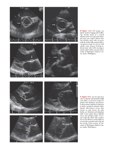
280 P A R T III / Assessment of Heart Disease
■ Figure 13-4 2-D images can
also be used for imaging valve leaflets.
Top: Systolic frame of a normal
trileaflet aortic valve (left) and a bicus-
pid aortic valve (right). Bottom: Nor-
mal trileaflet aortic valve in systole
with the leaflets fully opened (left); a
comparison image of a patient with
calcific aortic stenosis showing re-
duced aortic valve leaflet opening in
t
t
systole (right). (Echo courtesy of Uni-
versity of Washington Medical Cen-
ter, Seattle, Washington.)
■ Figure 13-5 Top: Normal mitral
valve in diastole with the leaflets fully
open (left); a comparison image of a
patient with rheumatic mitral steno-
sis demonstrates significant reduction
t
t
in leaflet opening in diastole (right).
Bottom: Myxomatous mitral valve
disease. In early systole, the valve
leaflets are thickened and redundant
t
t
(left). In late systole, there is bileaflet
mitral valve prolapse (right). The ar-
t
t
row marks the mitral valve annulus.
Notice the difference of the leaflets to
the annulus compared with early and
late systole. (Echo courtesy of Uni-
versity of Washington Medical Cen-
ter, Seattle, Washington.)