
Page 307 - Cardiac Nursing
P. 307
ara
g
g
ara
6/2
6/2
ara
3
xd
xd
3
0/0
0/0
3
M
g
2 A
M
Pa
Pa
M
1
009
009
1
2 A
0:4
0:4
p
p
p
A
83
83
A
t
LWBK340-c13_
LWB
LWB K34 0-c 13_ pp277-290.qxd 30/06/2009 10:42 AM Page 283 Aptara
K34
t
13_
0-c
7-2
7-2
q
90.
q
q
90.
e 2
p
p
27
27
e 2
C HAPTER 1 3 / Echocardiography 283
Diastole Systole
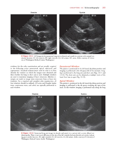
■ Figure 13-11 2-D images in the parasternal long axis in diastole and systole in a patient with normal ven-
tricular function. LV, left ventricle; RV, right ventricle; LA, left atrium; AO, aorta. (Echo courtesy of Univer-
sity of Washington Medical Center, Washington.)
windows for the echo examination and are usually acquired Parasternal Window
in the following order: parasternal, apical, subcostal, and The patient is positioned in the left lateral decubitus position and
suprasternal. Standard nomenclature used in echo is to first imaging is performed in the rib spaces left of the sternum. Imag-
identify the window in which the images were obtained and ing is performed in the long axis and short axis (Figs. 13-11 and
then whether the long or short axis is used. Multiple windows 13-12). The short axis can be obtained at multiple levels of the
are used to maximize imaging of heart structures, likewise at- heart from base to apex (Fig. 13-13).
tempts are made to image most structures from at least two
windows. For a standard and complete echo examination, all Apical Window
four windows should be utilized. 2-D echo and Doppler (pulse The patient is positioned in the left lateral decubitus position and
wave, continuous wave, and color) are typically performed in imaging is performed in the rib spaces overlying the apex of the
each window. heart. In this window, imaging is performed only along the long
Diastole Systole
■ Figure 13-12 Parasternal long axis images in diastole and systole in a patient with a severe dilated car-
diomyopathy. There is not much difference in the size of the left ventricle between diastole and systole, indicat-
ing poor systolic function. RV, right ventricle; LV, left ventricle; LA, left atrium. (Echo courtesy of University of
Washington Medical Center, Seattle, Washington.)