
Page 306 - Cardiac Nursing
P. 306
g
t
t
Pa
6/2
6/2
ara
e 2
0/0
0/0
e 2
82
g
g
0:4
0:4
1
2 A
M
M
2 A
ara
009
009
ara
1
M
Pa
3
A
p
p
A
p
p
82
LWBK340-c13_
LWB
LWB K34 0-c 13_ pp277-290.qxd 30/06/2009 10:42 AM Page 282 Aptara
K34
p
13_
0-c
xd
q
q
3
3
xd
q
7-2
27
27
7-2
90.
90.
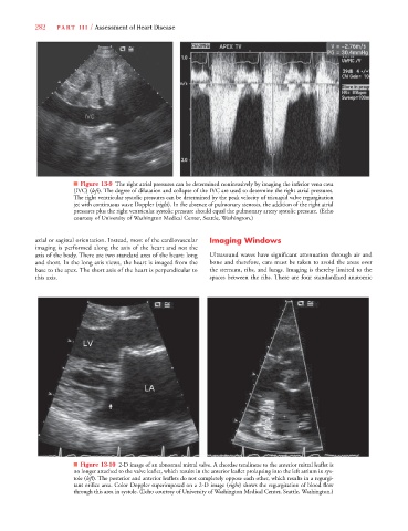
282 P A R T III / Assessment of Heart Disease
■ Figure 13-9 The right atrial pressures can be determined noninvasively by imaging the inferior vena cava
(IVC) (left). The degree of dilatation and collapse of the IVC are used to determine the right atrial pressures.
t
t
The right ventricular systolic pressures can be determined by the peak velocity of tricuspid valve regurgitation
jet with continuous wave Doppler (right). In the absence of pulmonary stenosis, the addition of the right atrial
t
t
pressures plus the right ventricular systolic pressure should equal the pulmonary artery systolic pressure. (Echo
courtesy of University of Washington Medical Center, Seattle, Washington.)
axial or sagittal orientation. Instead, most of the cardiovascular Imaging Windows
imaging is performed along the axis of the heart and not the
axis of the body. There are two standard axes of the heart: long Ultrasound waves have significant attenuation through air and
and short. In the long axis views, the heart is imaged from the bone and therefore, care must be taken to avoid the areas over
base to the apex. The short axis of the heart is perpendicular to the sternum, ribs, and lungs. Imaging is thereby limited to the
this axis. spaces between the ribs. There are four standardized anatomic
■ Figure 13-10 2-D image of an abnormal mitral valve. A chordae tendineae to the anterior mitral leaflet is
no longer attached to the valve leaflet, which results in the anterior leaflet prolapsing into the left atrium in sys-
tole (left). The posterior and anterior leaflets do not completely oppose each other, which results in a regurgi-
t
t
t
t
tant orifice area. Color Doppler superimposed on a 2-D image (right) shows the regurgitation of blood flow
through this area in systole. (Echo courtesy of University of Washington Medical Center, Seattle, Washington.)