
Page 329 - Cardiac Nursing
P. 329
CHAPTER 15 / Electrocardiography 305
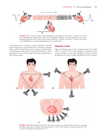
n Figure 15-7 A strip of cardiac muscle depolarizing in the direction of the arrow. A positive electrode at
B sees depolarization coming toward it and records an upright deflection. A positive electrode at A sees depo-
larization going away from it and records a negative deflection. A positive electrode at C records a flat line be-
cause depolarization is traveling perpendicular to the electrode’s view.
toward the left arm is recorded as a negative deflection. The right Unipolar Leads
leg electrode serves as a ground and does not contribute to the sig-
nals recorded. The electrical sum of the voltages in the three bipo- Figure 15-8B illustrates the three unipolar frontal plane leads,
lar frontal plane leads equals zero potential and forms a virtual aVR, aVL, and aVF. The camera represents the location of the
ground in the center of the triangle used by the unipolar leads as positive electrode: on the right shoulder for aVR, on the left
their reference point. shoulder for aVL, and at the foot (left leg) for aVF. The “negative
n Figure 15-8 The 12 leads of the ECG. The camera represents the location of the positive, or recording,
electrode in each lead. (A) Bipolar frontal plane leads I, II, and III. (B) Unipolar frontal plane leads aVR, aVL,
and aVF. (C) Unipolar precordial leads V 1 –V 6 .