
Page 332 - Cardiac Nursing
P. 332
1
0:3
1
/09
/09
0:3
M
Pa
M
0 P
0 P
q
q
q
32.
32.
xd
/29
/29
6
xd
6
Pa
ara
ara
t
p
t
a
c.
c.
In
a
In
e 3
e 3
g
g
g
08
p
p
A
08
A
K34
0-c
LWB
K34
0-c
30
30
15_
15_
LWB
0-3
0-3
LWBK340-c15_ p p pp300-332.qxd 6/29/09 10:30 PM Page 308 Aptara Inc.
308 P A R T III / Assessment of Heart Disease
a aVR
aVR
V
I I I I I aVR V V V V V V 1 1 1 1 V V V V V V V 4 4 4 4 4
R
VR
R
V
R
II II II
V
aVL
V
a a aVL
aVL V V V V V V 2 2 2 2 2 V V V V V V V 5 5 5 5 5
aVL
L
aVF
F
aVF
a a aVF
V
F
V
VF
IIII I III III III
I
V V V V V V 3 3 3 3 3 V V V V V V V 6 6 6 6 6
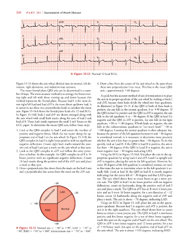
■ Figure 15-12 Normal 12-lead ECG.
Figure 15-13 shows the axis wheel divided into its normal, left de- 4. Draw a line from the center of the axis wheel to the spot where
viation, right deviation, and indeterminate sections. these two perpendicular lines meet. This line is the mean QRS
The mean frontal plane QRS axis can be determined in a num- axis—approximately
60 degrees.
ber of ways. The most accurate method is to average the forces mov- A quick but less accurate method of axis determination is to place
ing right and left with those moving up and down because this the axis in its proper quadrant of the axis wheel by looking at leads I
method represents the frontal plane. Because lead I is the most di- and aVF, because these leads divide the wheel into four quadrants.
rect right/left lead and lead aVF is the most direct up/down lead, it As illustrated in Figure 15-15, if the QRS in both of these leads is
is easiest to use these two perpendicular leads to calculate the mean positive, the axis falls in the normal quadrant, 0 to
90 degrees. If
4
4
axis. Figure 15-14A shows the frontal plane leads of a 12-lead ECG. the QRS in lead I is positive and the QRS in aVF is negative, the axis
In Figure 15-14B, leads I and aVF are shown enlarged along with falls in the left quadrant, 0 to 90 degrees. If the QRS in lead I is
the axis wheel with small hash marks along the axes of lead I and negative and the QRS in aVF is positive, the axis falls in the right
lead aVF. These hash marks represent the small 1-mV boxes on the quadrant,
90 to
180 degrees. If both leads are negative, the axis
ECG paper. To determine the mean QRS axis, follow these steps:
falls in the indeterminate quadrant or “no-man’s land,” 90 to
1. Look at the QRS complex in lead I and count the number of 180 degrees. Locating the correct quadrant is often adequate but,
positive and negative boxes. Mark the net vector along the ap- because the portion of the left quadrant between 0 and 30 degrees
propriate end of lead I on the axis wheel. In Figure 15-14B, the is considered normal, it is necessary to determine more precisely
QRS complex in lead I is eight boxes positive with no significant whether the axis is less than or greater than 30 degrees. To do this
negative deflections. Count eight hash marks toward the posi- quickly, look at Lead II: if the QRS in lead II is positive, the axis is
tive end of lead I and put a mark on the axis wheel at that spot. less than 30 degrees; if the QRS in Lead II is negative, the axis is
2. Look at the QRS complex in aVF and follow the same proce- more negative than 30 degrees indicating LAD.
dure as before. In this example, the QRS complex in aVF is 14 Using the ECG in Figure 15-16A, first place the axis in the ap-
6
6
boxes positive with no significant negative deflections. Count propriate quadrant by using leads I and aVF. Lead I is upright and
14 hash marks along the positive end of the aVF axis and place aVF is negative, placing the axis in the left quadrant. However, be-
a mark at that spot. cause 30 degrees of the left quadrant is considered normal, we need
3. Draw a perpendicular line down from the mark on the lead I axis to fine-tune the axis to determine where in the left quadrant it ac-
and a perpendicular line across from the mark on the aVF axis. tually falls. Look at lead II: the QRS in lead II is mostly negative
indicating that the axis is left of 30 degrees and that LAD is pres-
ent. The axis wheel shows how to count boxes to get a more pre-
cise axis. The QRS in lead I is six boxes positive with no negative
90
deflections; count six hashmarks along the positive end of lead I
axis and place a mark. The QRS in aVF has an R wave 4 boxes pos-
Indeterminate LAD 30 itive and an S wave 16 boxes negative, for a net direction of 12
boxes; count 12 hashmarks along the negative end of aVF and
Using the ECG in Figure 15-16B, place the axis in the appro-
180 place a mark. The axis is about 70 degrees, indicating LAD.
priate quadrant. Because lead I is negative and aVF is positive, the
Normal
RAD axis is in the right quadrant. The axis wheel shows how to count
boxes to obtain a more precise axis. The QRS in lead I is two boxes
positive and five boxes negative for a net of three boxes negative;
mark this spot on the negative end of lead I on the axis wheel. The
90 QRS in aVF is two boxes negative and 12 boxes positive for a net
■ Figure 15-13 Normal axis 30 to
90 , LAD 31 to of
10 boxes; mark this spot on the positive end of lead aVF on
90 , RAD
91 to
180 , indeterminate axis 91 to 180 . the axis wheel. The axis is about
110 degrees, indicating RAD.