
Page 333 - Cardiac Nursing
P. 333
M
Pa
0 P
M
Pa
g
e 3
g
g
/09
/09
/29
/29
1
0:3
0 P
1
0:3
ara
a
t
ara
a
c.
c.
In
In
09
A
e 3
09
A
p
t
p
p
p
p
15_
30
0-3
0-3
30
15_
LWB
LWB
LWBK340-c15_ pp300-332.qxd 6/29/09 10:30 PM Page 309 Aptara Inc.
K34
0-c
0-c
K34
32.
q
q
q
xd
xd
6
6
32.
C HAPTER 1 5 / Electrocardiography 309
I I I I aV R
R
R R R R R R R R
R
V
aVV
aV
V
V
V
a a a a a a a a a a a a
V
VR
R
V
R
V
V V V V
VRR
V
R
R
VR
VR
V V V V
V
V
V
V
V
V
V
VL
VL
V
a a a a a a a a a a a a
aVV
L L L L
L
aV
VL
I I I I I I I I aV L
L
VLL
a a a a a a a a a a a a a a a
aV
aVV
V
V
V
V
V
F F F F
F
F
V
VFF
VF
VF
VF
I I I I I I I I I I I I aV F
V
V
V V V V
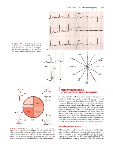
■ Figure 15-14 Calculating the mean
QRS axis. (A) The six frontal plane leads of
an ECG. (B) Lead I and lead aVF enlarged.
See text for instructions on calculating the A
axis using leads I and aVF on the axis wheel.
90
I I 120
8 8 8 8 60
150 30
AVR AVL
V
V
AV F
180 I 0
A A
AVF
F
F
14
1 1
14 4 4
150
30
III
120 II
AVF
60
90
I I I I I I I I
INTRAVENTRICULAR
I I I CONDUCTION ABNORMALITIES
D A
The intraventricular conduction system consists of the right bundle
branch and the left main bundle branch, which fans out into septal
F F F F F F F F F F F F
F F F F F F F F F F F F fascicles, an anterior fascicle, and a posterior fascicle. There are nu-
LAD merous individual anatomic variations, but the intraventricular con-
I I I I I I I I I I I I I I I I I I I I I I I I I I I I I I I I I I I I I I I I I I I I I I I I I I I I I I I I I I I I I I I Indeterminate
duction system is generally regarded to consist of three major fasci-
I cles that diverge from the bundle of His: (1) the right bundle branch,
(2) the anterior division of the left bundle branch (left anterior fas-
cicle), and (3) the posterior division of the left bundle branch (left
RAD Normal
9
I I I posterior fascicle ; Fig. 15-17). Block may occur in any part of this
I I I conduction system. Monofascicular block involves block in only one
of the three major fascicles. The term bifascicular block is most com-
F F F F F F F F F C AVF B monly used to describe the combination of RBBB and either LAFB
F F F F F F F F F F F F or LPFB. Trifascicular block means block in all three major divisions.
Bundle-Branch Block
■ Figure 15-15 The four quadrants of the axis wheel. (A) If the
QRS in lead I is positive and the QRS in aVF is negative, the axis is When one of the bundle branches is blocked, the ventricles depo-
in the left quadrant. (B) If the QRS is positive in both leads I and larize asynchronously. Bundle-branch block is characterized by a
aVF, the axis is normal. (C) If the QRS in lead I is negative and the delay of excitation to one ventricle and abnormal spread of elec-
QRS in aVF is positive, the axis is in the right quadrant. (D) If the trical activity through the ventricle whose bundle is blocked. This
QRS is negative in both leads I and aVF, the axis is indeterminate. delayed conduction results in widening of the QRS complex to