
Page 335 - Cardiac Nursing
P. 335
/09
/09
c.
1
0:3
0:3
1
/29
/29
6
a
c.
In
In
0 P
g
g
g
e 3
11
11
e 3
M
A
0 P
M
Pa
Pa
A
30
30
p
p
p
32.
0-3
0-3
LWBK340-c15_
K34
LWB K34 0-c 15_ pp300-332.qxd 6/29/09 10:30 PM Page 311 Aptara Inc.
LWB
p
p
0-c
15_
6
xd
xd
q
ara
q
q
32.
t
t
a
ara
C HAPTER 1 5 / Electrocardiography 311
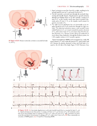
1. Septal activation occurs first from left to right, resulting in the
normal small R wave in V 1 and small Q wave in V 6 .
2.The left ventricle is activated next through the normally func-
tioning left bundle branch. Depolarization spreads normally
2 through the Purkinje fibers in the left ventricle, causing an S
V 6 wave in V 1 as the impulse travels away from its positive elec-
2 2 2 2 trode and an R wave in V 6 as the impulse travels toward the
1 1 1 1 1 positive electrode in V 6 .
3. The right ventricle depolarizes late and abnormally as the im-
1 pulse spreads by cell-to-cell conduction through the right ven-
tricle. This abnormal activation causes a wide second R wave
1
(called Rprime) in V 1 as it travels toward the positive electrode
in V 1 , and a wide S wave in V 6 as it travels away from the pos-
itive electrode in V 6 . Because muscle cell-to-cell conduction is
much slower than conduction through the Purkinje system, the
V 1 QRS complex widens to 0.12 second or greater.
2
Typical uncomplicated RBBB can be recognized by a wide rSR
■ Figure 15-18 Normal ventricular activation as recorded by leads prime pattern in V 1 and a wide qRs pattern in V 6 and in leads I and
V V
V 1 and V 6 .
aVL, because the positive electrode in these two limb leads is lo-
cated on the left side of the body. Figure 15-20B illustrates three
2
V 6
2 2 2
I I I
1 1 1 1 1
3 3 3 3 3 3 3 1
3
3
1
1
A 2 B
R R R R R R R R R
R
a a a a a a a a
aV
R
aVVVR
V
V
V V V V V VR
I I I I I aV R V V V V V V V V V V V V V V V 1 1 1 1 V V V V V V V V V V V 4 4 4 4
V
VR
V
VL
V
V
V
V
V
V
V
a a aVL
a a aVL
III I III I III I III I II II II aVL L L L L L L L L V V V V V V V V V V V V V V V V V V V V V V V 2 2 2 2 2 2 2 2 V V V V V V V V V V V V V V V V V V V 5 5 5 5 5 5 5 5
aVL
V
V
a a aVL
V
VL
V
V
aVF
V
V
V
V
III III III III III III a a aVF V V V V V V V V V V V V V V V V V V 3 3 3 3 3 3 3 3 V V V V V V V V V V V V V V V 6 6 6 6 6 6 6 6
aVFF
a a aVF
a a aVF
F
F
F
VF
V
V
V
V
F
F
C
■ Figure 15-19 (A) Ventricular depolarization with right bundle branch block as recorded by leads V 1 and
V 6 . Septal activation occurs first (arrow 1) causing an R wave in V 1 and Q wave in V 6 ; left ventricular activa-
tion occurs second (large arrow 2) causing an S wave in V 1 and an R wave in V 6 ; right ventricular activation
occurs last and slowly (curved arrows 3) causing an R’ in V 1 and a wide S wave in V 6 . (B) Three commonly seen
variations of RBBB pattern. (C) 12-lead ECG illustrating RBBB.